Polipoză de sinus frontal operată prin abord endoscopic transcranian
Transcranian endoscopic approach in a frontal polyposis
Abstract
We present the case of a 62-year-old patient with a large bilateral frontal sinus tumor, with severe headache and a partial destruction of the left lamina papyracea and of the posterior wall of the right frontal sinus. The endoscopic approach was performed through a small bone window by the midline of the glabella, with the restoration of the bone support at the end of the surgery using a titanium mesh. Although the surgical staff has a lot of experience in doing the endoscopic transnasal approach, there are some cases where the transnasal route cannot safely solve certain pathologies. This is the reason why the authors wish to illustrate the need of using an approach that will ensure a proper management of any kind of complications that can occur during surgery, specific to these tumors (bleeding, cerebrospinal fluid leak). In this case, the transcranial endoscopic approach was the best solution.Keywords
frontal sinustumorexternal endoscopic approachRezumat
Prezentăm cazul unui pacient, în vârstă de 62 de ani, cu o formaţiune tumorală voluminoasă de sinus frontal bilateral, cu sindrom cefalalgic sever şi distrucţia parcelară a laminei papiracee stângi şi a peretelui posterior al sinusului frontal drept. Abordul endoscopic s-a efectuat printr-o fereastră intersprâncenoasă de mici dimensiuni, cu refacerea suportului osos la finalul intervenţiei utilizând o plasă din titan. Deşi echipa chirurgicală are o experienţă îndelungată în abordul transnazal endoscopic, există cazuri în care parcursul transnazal nu poate rezolva în deplină siguranţă anumite patologii. Autorii doresc să sublinieze necesitatea utilizării unui abord care să asigure managementul corespunzător al unor eventuale complicaţii intraoperatorii specifice acestor tumori (sângerări, fistulă de lichid cefalorahidian), iar în cazul de faţă abordul endoscopic transcranian a reprezentat cea mai bună soluţie.Cuvinte Cheie
sinus frontaltumorăabord endoscopic externA 62-year-old patient, S.I., came to our hospital with an intensive headache syndrome, treatment-resistant, which had been known for several years and had worsened in the last 2-3 weeks. Three years ago (May 2016), in another ENT service, the patient underwent a curative surgery for bilateral fronto-ethmoido-sphenoidal rhinosinusitis. According to the medical discharge presented by the patient, the frontal sinus approach used was of the Draf I kind, without exploring the frontal sinus cavity.
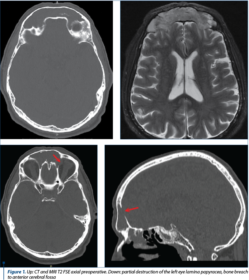
The CT/MRI scans on admission revealed the existence of a tumor-like formation, with intense gadolinium (hypercaptant), that had ballooned both frontal sinuses, and destroyed the intersinusal wall.
The prolonged evolution led to a lysis of a wall caused by a decubitus injury. This explained the intimate tumoral contact with the dura mater through an 8-mm bone breach in the posterior wall of the right frontal sinus.
Also, the tumor protruded through the left orbit by an erosion of the left lamina papyracea, with the appearance of a discrete inferior and external exophthalmia in the left eye. The MRI revealed that the dura mater was integral and the orbital periosteum was apparently free (Figure 1).
The apparent origin located at the posterior wall of the bilateral frontal sinus, the partial exposure of the dura mater and the destruction of the lamina papyracea with the penetration of the tumor into the left orbit led to the decision of an external endoscopic approach. We considered that a possible cerebrospinal fluid (CSF) leak at the posterior wall of the right frontal sinus, as well as an important intraoperative bleeding couldn’t be managed properly by a transnasal approach.
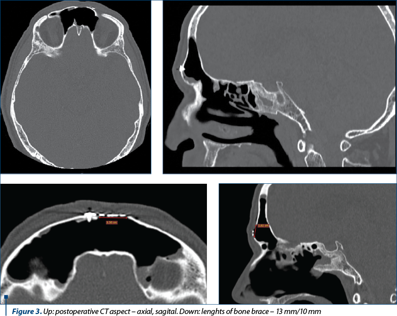
We underwent the surgery using general anesthesia with OT intubation. Although the tumor was large (horizontal diameter: 82 mm, antero-posterior: 18 mm, vertical: 43 mm), we decided that the approach should be performed by the midline of the glabella, through a bone window that would allow the access of a telescope with a 4 mm diameter plus two other tools (suction tube + endoscopic forceps or a 2-mm microdebrider). Skin incision: 20 mm, bone fenestration with 13 mm horizontal diameter and 10 mm vertical diameter.
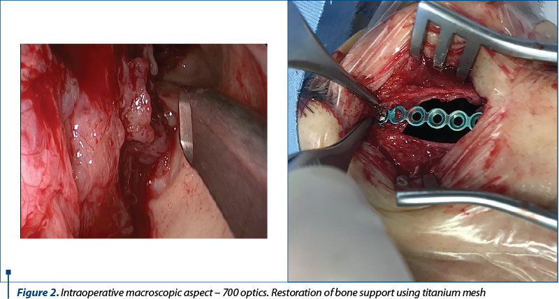
A hard tumor was shown upon palpation, with a macroscopic aspect of an inverted papilloma, well-vascularized, that occupied both frontal sinuses and dived through the nasofrontal ducts in the anterior ethmoidal cells. The apparent origin of the tumor was located at the junction between the posterior wall of the left frontal sinus and the intersinusal septum, in the upper floor of the sinus cavity.
Although the approach was carried out through a small hole, using the 300, 700 and 900 angled optics and the angled tools (Heuwieser forceps of 700 and 900, angled suction tube and a monopolar malleable suction cautery), we achieved a full-macroscopic abaltion using “piece-meal” method. There was a partial exposure of the dura mater and the left orbital periosteum, but there was no signs of penetration at their level.
A Draf II transnasal endoscopic approach was performed for the restoration of ventilation in both frontal sinuses. Radiofrequency hemostasis – fulfuration 10 W. Efficient hemostasis; did not require nasal package. The integrity of the bone support was restored using a titanium mesh fixed with biocompatible screws (Figure 2). Intradermal skin suture (Vicryl 5.0).
A CT scan using a contrast substance was performed at the end of the surgery (Figure 3). It confirmed the complete macroscopic resection of the tumor and a wide ventilation of both frontal sinuses.
Although, macroscopically, the appearance suggested an inverted papilloma, the IHC examination of the resected piece described tissue fragments covered by a respiratory type epithelium, with edema-like stroma with abundant chronic inflammatory infiltrate adding eosinophils and normal-looking glandular structures. IHC revealed the diagnosis of a glandular subtype of sinus inflammatory polyp.
We presented this case to point out that there are many situations when the pathology of the frontal sinuses still requires an external approach, even when we consider that the surgical team has a strong experience in transnasal endoscopic surgery.
The peculiarity of the case was the small bone-window through which the tumor was resected, the advantage we had being given by the usage of optics and angular tools that made the resection complete and safe for the patient.
CSF leaks, the frontal sinus osteomas that exceed the diameter of the nasofrontal duct and large solid tumors can be safely managed for the patient through a minimally invasive transcranial endoscopic approach that is lesion-centered.
Note: This article does not contain any references, since it describes a strictly personal experience of the authors.
Conflict of interests: The authors declare no conflict of interests.
Bibliografie

The First National Congress of the young Romanian Society of Audiology and Communication Pathology
Sebastian Cozma
Between the 12th and 14th of September 2019, we had the joy of meeting in a special academic and cultural setting offered by the Palace of Culture in Iaşi, on the occasion of the First National Congre...
Aspecte particulare ale anatomiei etmoidale pe imagini CT
Alexandra Gheorghe, Silviu Crăc, A. Panfiloiu, Vlad Andrei Budu
Anatomia ilustrată pe descoperirile imagistice reprezintă baza înţelegerii structurilor normale şi patologice rinosinuzale. Actualmente, radiologia convenţională a fost înlocuită de imagistica CT, noul standard de au...
Utilizarea pivoturilor endodontice în restaurările odontale armate pe dinţi devitali
Irina-Maria Gheorghiu, Paula Perlea, Claudiu-Gabriel Ciolan, Loredana Mitran, Mihai Mitran, Alexandru Iliescu
În situaţiile clinice în care se impune restaurarea distrucţiilor coronare masive, este obligatorie utilizarea mijloacelor suplimentare de retenţie ce pot asigura succesul şi menţinerea restaur...
Carcinomul de vestibul nazal – comentarii pe baza unui caz clinic
Mihai Tușaliu, Lavinia-Georgiana Ilinca, Iulia Tiţă
Întâlnit la mai puţin de 1% din totalitatea neoplasmelor capului şi gâtului, carcinomul cu localizare la nivelul vestibulului nazal este o patologie rară în practica ORL, cu un tablou clinic nespecific. Mijloacele te...
Frontal sinus osteoma – case report
Celesta Drăgulescu, M. Chițac, A. Weisman, M. Condrat, X. Dolghii, Emanuela Onisâi, M. Vasilca
Osteomul reprezintă o tumoră benignă a sinusurilor paranazale, afectând cu predilecţie sinusul frontal. Are o evoluţie lentă şi silenţioasă, fiind descoperit adesea accidental prin examenul CT efectuat pentru diagnosti...