Din aceeași categorie

Opinii şi recomandări curente pentru utilizarea meşei în chirurgia ginecologică – review al literaturii
Introduction
Uterine myomas are smooth muscle benign tumors, with a prevalence of 2-10% during pregnancy(1-3). The development of uterine fibroids is influenced by genetic factors and the hormonal levels of estrogen and progesterone(4-7). Therefore, their growth is expected during pregnancy, but most of them do not change significantly in size during pregnancy(6,8,9). It is a common belief that uterine myomas increase in size during pregnancy, but this happens in rare cases(10). The growth of uterine myomas in pregnancy cannot be predicted(11). Still, some studies found that, during the first and the second trimesters of pregnancy, the fibroids may remain unchanged, increase or even decrease in volume, or remain at the same size in the third trimester(12).
The presence of fibroids in pregnancy is usually asymptomatic, but they can be associated with possible complications(6).
The most common symptoms of uterine myomas in pregnancy, according to Spyropoulou et al. (2020), are abdominal pain, fever, abdominal heaviness, vomiting, constipation and vaginal bleeding. Other symptoms related to the presence of uterine fibroids might be urine retention, respiratory discomfort, uterine contractions and hydronephrosis(2).
The most common symptom is pain(13,14), caused by pressure of the fibroid itself or by torsion of a pedunculated myoma(6). The pelvic pain has been described to be more severe in posterior-located fibroids, larger than 30 mm in diameter than anterior-located ones(14).
The syndrome of painful myoma describes the association of severe abdominal pain, nausea, vomiting and fever(15,16), and it is considered the major complication of myomas in pregnancy(17). The common causes of painful myoma syndrome are red or carneous degeneration(11), torsion of the pedicle, and infection or necrosis(18).
Leiomyomas can suffer several degenerative changes, such as hyaline degeneration or red or carneous degeneration, which is considered specific but not exclusively pregnancy-related. Often, they are causing pain and fever(16,19).
The miscarriage rate in pregnancy associated with submucosa myomas is 14%, almost double compared to the pregnancies without fibroids(20-22). However, a systematic review and meta-analysis conducted by Sundermann et al. (2017), including more than 20,000 women, did not find any association between spontaneous abortion and uterine myomas(23).
When the uterine myomas have an increased size, they may cause pollakiuria by irritating the urinary bladder, along with an increased blood flow in pregnancy, retention of urine by compressing the bladder neck, or even urinary tract infections due to intermittent urinary retention caused by partial obstruction(6). Also, compression of large posterior myomas on the rectum may cause chronic constipation(24). Another, yet very rare, complication of posterior uterine wall myomas associated with a retroverted uterus is the pelvic incarceration of the pregnant uterus(6,25).
Premature rupture of membranes was significantly increased in pregnancies associated with fibroids, in a small study(26). Still, other studies do not support this finding(27).
The presence of uterine myomas may increase the risk of premature uterine contractions and premature birth(2,28). A retrospective cohort study performed in France, including almost 20,000 pregnant women, from which 301 had uterine myomas, has reported a 2.5-fold increased rate of preterm birth in pregnancies associated with uterine myomas(29).
The risk of placental abruption has been reported in some studies, occurring most in submucous and retroplacental fibroids having a diameter above 7-8 cm(29-32). Other studies found no correlation between placental abruption and fibroid(8,12).
Post-partum hemorrhage is more common in pregnancies associated with uterine myomas than in uncomplicated pregnancies(27,29). It is assumed that overdistension of the uterus due to the presence of fibroid predisposes to uterine atony and bleeding(33,34).
A distorted uterine cavity, related to fibroids, can cause fetal malpresentation(29,34-36), especially in breech cases(37). In rare cases, the fetus may be affected by the presence of the fibroid, causing fetal deformation (abnormal position of limbs, congenital torticollis and head deformities)(6,33).
After delivery, the fibroids may regress spontaneously or may complicate with torsion in the case of pedunculated fibroids or ischemic degeneration in the case of submucosal fibroids, providing an excellent environment for anaerobic bacterial culture and, therefore, puerperal fever and tachycardia associated with intense pelvic pain(6).
The mode of delivery in cases associated with uterine myomas is influenced by the number, size and location of the myomas. In large myomas cases, there is a higher incidence of caesarean delivery(37); however, the presence of uterine myomas does not contraindicate the trial of labor unless the myomas obstruct the birth canal. Therefore, patients with fibroids larger than 5 cm and located in the inferior uterine segment have an increased rate of caesarian delivery(29,38-41).
The first-line treatment of uterine myomas in pregnancy is represented by conservative management of the symptoms, with symptomatic treatment and bed rest. When, despite maximal analgesics and prostaglandin synthesis inhibitors, the abdominal pain persists, it becomes more severe in large or rapidly growing myomas, large or medium myomas located in the lower uterine segment, or deforming the placental site, the conservative treatment fails and the surgical treatment is recommended(16).
A systematic review of myomectomy in pregnancy reports a small number of complications. In this review, most fibroids were subserous and subserous pedunculated, located at the uterine fundus, with no contact with the placenta. In cases of single and multiple myomectomies, fibroids removed were intramural and subserous located at the uterine fundus. This review includes 76 myomectomies by laparotomy, 15 by laparoscopy, four by vaginal approach, and one case of operative hysteroscopy(2).
Antibiotic prophylaxis and tocolysis before and during the surgery are recommended for preoperative care. Some authors consider medial laparotomy the best approach(15,16,18,24,42-73). Other authors advocate for a laparoscopic approach(1,74-83). Vaginal surgery is a valid option for submucosal pediculated myomas located in the cervical canal(49,84-86).
The main bias in myomectomy in pregnant women is the increased risk of massive bleeding. Outside of pregnancy, controlled hypotensive anesthesia, tourniquets and local injections of vasoconstrictive agents represent valid options to prevent intraoperative hemorrhage, but in pregnancy those are not allowed. To reduce the risk of massive bleeding, Suwandinata et al. (2008) described a technique of interrupted sutures placed around the myoma to secure the blood vessels encircling it before performing myomectomy(16).
Spyropoulou et al. (2020)(2), in a systematic review of 97 cases of myomectomy in pregnancy, reported surgery complications in less than 10% of the cases, as follows: moderate vaginal bleeding(24), cervical shortening with cervical positive culture(45), miscarriage(15,42,45,57,70,84), rupture of membrane shortly after the surgery(84), hematoma of the uterine scar(49), uterine rupture following myometrial necrosis and abscess(77), and a case with purulent chorioamnionitis(42).
Routine myomectomy during caesarean section is not recommended, due to an increased risk of massive bleeding, but it might be feasible and safe in tiny and pedunculated myomas(87).
Case report
We present the case of a 31-year-old primigravida presenting with severe abdominal pain, pollakiuria and constipation. The patient did not report a relevant medical personal or familial history.
The gynecological exam revealed an enlarged uterus for gestational age, with the uterine fundus elevated to the umbilical scar.
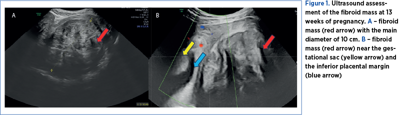
The ultrasound examination revealed a 13-week pregnancy, with no ultrasound signs of fetal structural abnormalities, and several fibroids, in contact with each other, developed as one fibroid mass, intramural and subserous, in the lower uterine segment and into both right and left parametria, with a diameter of 100/95/87 mm and a volume of 826 cm3 (Figure 1A). The placental attachment was on the anterior uterine wall, with no contact with the fibroid mass, but in close proximity – 3 mm (Figure 1B).
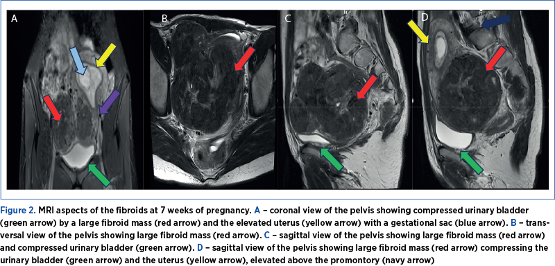
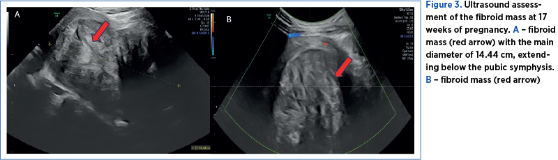
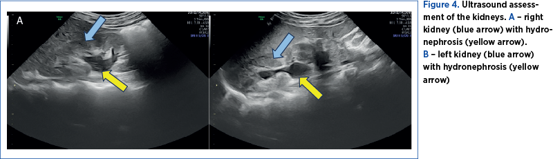
The first pregnancy ultrasound assessment, performed at 7 gestational weeks, described uterine fibroids, and a pelvic magnetic resonance imaging (MRI) was recommended. The results of the pelvic MRI confirmed the ultrasound report. Moreover, the MRI report provided additional information about the development of the myomas, submucosal and sub-serosal, with more than 50% intramural development, and their mass effect on the uterine cavity, bladder and rectum (Figure 2 A-D).At first, we recommended a urinary test for infections, and analgetic and prostaglandin synthesis inhibitors treatment for the management of pelvic pain. During the following weeks, the symptoms progressed, restricting daily activities as the fibroid volume increased, reaching a diameter of 150/90/95 mm, with an almost double volume (1425 cm3). Also, their ultrasound aspect slightly changed to a lower echoic heterogenous aspect (Figure 3 A, B). The severe pelvic pain, along with the increased size of the fibroid and the suggestive ultrasound aspect raised the suspicion of fibroid degeneration. The increased size of the myomas caused yet another symptomatic complication – bilateral ureteral compression with hydronephrosis (Figure 4 A, B).
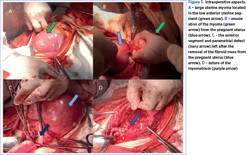
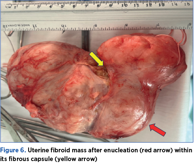
Therefore, at 17 weeks of pregnancy, due to severe abdominal pain, interfering with daily activities, not responding to analgesics, the rapidly growing myomas and the ultrasound aspect, we decided to perform a myomectomy by midline laparotomy approach. We found a 150/150/90 mm fibromatous mass developed in the inferior uterine segment, the left parametrium and the right parametrium near the right uterine artery, and we removed it by enucleation (Figure 5 A, B). The removal of the fibroid mass was uneventful, as we avoided injury to the right uterine artery by performing the mass dissection within its capsule (Figure 6). The myometrium was sutured in two layers using resorbable sutures (Figure 5 C, D). Overall, the surgery was uneventful. The postoperative recovery went well, with antibiotics prophylaxis, low-molecular-weight heparin, progesterone, tocolytic and antispastic treatment, and analgesics for managing postoperative pain.
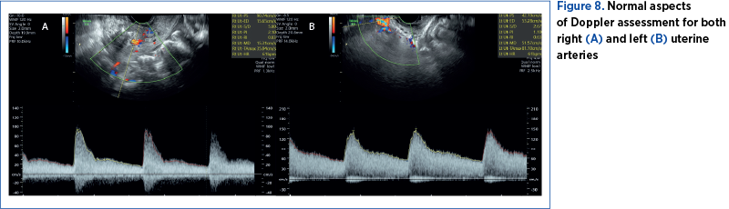
The patient was discharged after four days. At that time, the pregnancy evaluation revealed normal ultrasound relations. The uterine scar was visualized on ultrasound with a normal aspect and no signs of hematomas (Figure 7 A, B). The uterine artery Doppler interrogation showed indices within the normal range (Figure 8 A, B). We also calculated a normal amniotic fluid index (Figure 7C).
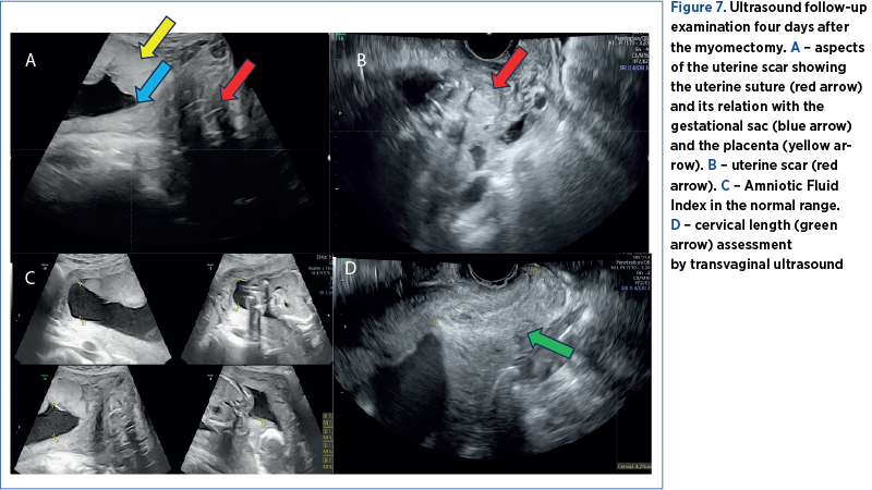
The cervical length was difficult to assess before the surgery due to the position of the fibroid mass, which distorted the uterine anatomy, pushing the cervix in a posterior-lateral position, difficult to assess by ultrasound or by clinical examination, since the myomatous mass extended till the vaginal introitus. Therefore, we only assessed the cervical length after the surgery in several examinations and maintained above the normal cutoff value (Figure 7D).
The patient was monitored weekly. A detailed second-trimester anomaly scan was performed at 22 weeks. It showed no signs of fetal structural abnormalities, an estimated fetal weight at percentile 30, and a pulsatility index within normal ranges for both uterine and umbilical arteries.
The histopathological exam revealed edematous degeneration of the myomas, as we suspected on ultrasound.
Overall, the mother and the fetus had a good follow-up assessment after myomectomy. The last follow-up exam performed at 32 weeks showed normal growth, with an estimated fetal weight at the 55th percentile.
Discussion
Uterine myomas have a relatively high incidence in pregnant women, at about 10%(1-3). It is well known that genetics and hormonal levels of estrogen and progesterone influence the development and growth of myomas(4-7). Our patient has no familial history of uterine myomas or other gynecological-related conditions. Regarding the hormonal influence on fibroid growth, studies show that the high levels of estrogens and progesterone in pregnancy do not usually determine a significant development of uterine myomas, most of them remaining at the same size during the pregnancy. However, in our case, some of them may have a rapid growth(12), where the fibroid mass almost doubled the volume in four weeks.
Fibroids in pregnancy are usually asymptomatic, but their presence may influence the pregnancy outcome, depending on the size, location and associated symptoms(6). The most common symptom is severe abdominal pain, accompanied, in large myomas, by compression symptoms(2). In our case, along with the severe abdominal pain, the fibroid mass compressed both the bladder and the rectum, producing urinary symptoms such as pollakiuria and digestive symptoms such as constipation. The first line of treatment is conservative, with analgesics and prostaglandin synthesis inhibitors(16). Despite maximal analgesic and prostaglandin synthesis inhibitors treatment, the symptoms of our patient progressed. Furthermore, the compression due to the increasing size of the myoma added new symptoms, such as hydronephrosis. Along with the rapid growth of the myomas, we also noticed a change in the ultrasound aspect, suggesting the degeneration of the fibroid mass.
Ultrasound examination is the first-line imaging method to diagnose uterine myomas, being relatively cheap and available in every clinic(88,89). Magnetic resonance imaging is the most accurate imaging technique for detecting, localizing and characterizing large myomas. This investigation is considered safe in pregnancy, offering advantages over ultrasound in assessing myoma burden(89,90). In our case, MRI brought essential information about the location of the myomas, from G2 to G5, according to FIGO staging of uterine myomas(91). Their position regarding the gestational sac, the cervix, and the compression on the uterine cavity, urinary bladder and rectum were also better exposed with MRI.
The myomectomy in pregnancy can be performed by laparotomy, laparoscopy, and even vaginal surgery in selected cases(2). Our approach by laparotomy was guided by the increased size of the myomas, estimated at almost 15 centimeters. When performing a surgical procedure on a pregnant uterus, the manipulation must be gentle, to reduce the risk of additional pressure on the pregnant uterus. Increased vascularization in pregnancy increases even more the risk of intraoperative hemorrhage. Due to the sensitivity of the fetus to hypoxia, hypotensive controlled anesthetic techniques, use of tourniquets and local injections of vasoconstrictive agents are contraindicated(16). In the attempt to prevent massive intraoperative hemorrhage, a technique of interrupted sutures placed around the myoma to secure the blood vessels encircling was described(16). In our experience, a carefully performed myomectomy within the fibrous capsule of the myoma is considered safe, with minimal blood loss. The site and the size of the myoma may also represent a technique challenge. The fibroid mass of our patient was estimated to be around 15 cm, developed on the anterior uterine segment and the right parametrium, in close rapport with the right uterine artery. After the urinary bladder dissection and myomectomy, we chose to perform a double-layer suture of the myometrium. By keeping the dissection inside the capsule, along with a careful myometrial suture, we managed to avoid the uterine artery injury confirmed by normal Doppler spectrum on further examinations.
As recommended, antibiotic prophylaxis and tocolysis were administrated before surgery(16). Afterward, we continued the tocolytic treatment along with progesterone, antispastic treatment, low-molecular-weight heparin, and analgesics for the management of pain.
After avoiding intraoperative massive hemorrhage and pregnancy loss, we carefully monitored the uterine scar for hematoma. The scar had an uneventful evolution, with a normal aspect on ultrasound.
Further follow-ups of the pregnancy were made, with a favorable growth of the fetus.
The increased size of the myomas, blocking the birth canal, would have represented an absolute indication of caesarean delivery(37). Even after the myoma removal, vaginal delivery is not recommended, due to the risk of uterine rupture(92).
Conclusions
Myomectomy should be considered in pregnant women with symptomatic uterine fibroids, not responding to conservative management, large or rapidly growing, located in the lower uterine segment, or deforming the placental site.
The procedure may associate an increased rate of complications during pregnancy, but with the right preparation and experience, it is considered a safe procedure.
Conflict of interest: none declared
Financial support: none declared
This work is permanently accessible online free of charge and published under the CC-BY.