Maxilectomia maxilară medială pentru papilomul inversat
Endoscopic medial maxillectomy for inverted papilloma
Abstract
Medial maxillectomy represents the gold standard technique for surgical treatment of inverted papilloma, as it allows the proper exposure of the lateral nasal wall and maxillary sinus. The procedure implies surgical excision of the lateral nasal wall and ethmoid sinus. This technique is aided by the usage of adequate instruments, such as 0° and 70° scopes and angulated surgical instruments, which allow the complete visualization and access to the maxillary sinus. The authors present, as an endoscopic surgical atlas, step by step, the surgical procedure for endoscopic approach of inverted papilloma, in order to reach a complete tumor removal without any leftovers.
Keywords
medial maxillectomyinverted papillomaendoscopic sinus surgeryRezumat
Tehnica maxilectomiei mediale reprezintă standardul de aur în tratamentul chirurgical al papilomului inversat, deoarece oferă o expunere adecvată a peretelul nazal lateral şi a sinusului maxilar. Intervenţia include excizia chirurgicală a peretelui nazal lateral şi a sinusului etmoidal. Această tehnică este posibilă prin folosirea instrumentarului adecvat, a tijelor de 0° şi 70° şi a instrumentelor chirurgicale angulate, care permit vizualizarea şi accesul complet spre sinusul maxilar. Autorii prezintă, ca un atlas de chirurgie endoscopică, paşii acestei tehnici chirurgicale de abord endoscopic al papilomului inversat, în vederea exciziei complete a tumorii, fără nicio restanţă.
Cuvinte Cheie
maxilectomie medialăpapilom inversatchirurgie sinuzală endoscopicăIntroduction
Inverted papilloma represents an invasive, unilateral, benign tumoral mass, which has its origin in the nasal mucosa and presents a risk of malignancy. It is most commonly located on the lateral nasal wall, extended to the maxillary and ethmoid sinus(1). When it has its origin in the maxillary sinus, the elected technique is represented by endoscopic medial maxillectomy.
Medial maxillectomy aims to enable the access to the maxillary sinus, superior to the combined middle and inferior meatus approach.
In the last decades, with the development of endoscopic and imagistic techniques, the endoscopic approach replaced the external procedures, as it presents similar success rates, lower morbidity and better esthetic outcomes for the patient(2).
Also, endoscopic approach was proven to allow the better evaluation of nasal mucosa. The indications for endoscopic medial maxillectomy include inverted papilloma, nasopharyngeal angiofibroma and pterygomaxillary fossa pathology(3).
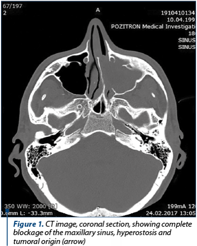
The diagnostic protocol depends on a computed tomography, which illustrates the extension and place of attachment of the tumor.
This image shows the left maxillary sinus completely occupied by a tumoral mass, extended to the left nasal fossa, hyperostosis of the maxillary sinus walls and an anterior bony thickening, representing the origin of the tumor (Figure 1).
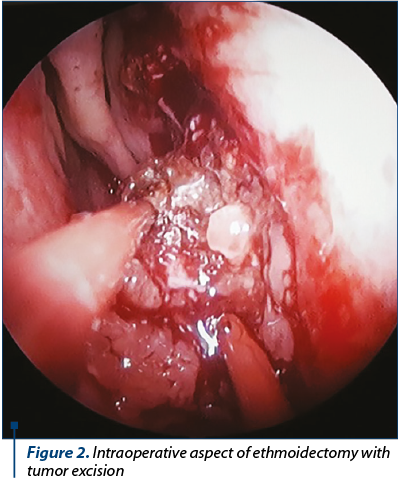
The first surgical step includes antero-posterior ethmoidectomy, with the excision of the tumor from this level and from the middle meatus. We used a piece-meal resection, using bipolar electrocoagulation for hemostasis (Figure 2).
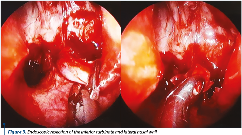
After ethmoidectomy and clearing of the middle meatus, the resection of the inferior turbinate and sinonasal wall is performed. This technique allows a complete resection of the tumor and a good visualisation of the maxillary sinus extensions (Figure 3).
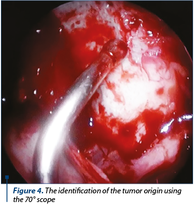
Medial maxillectomy offers an optimal access to the maxillary sinus, with the complete resection of the tumor and the identification of the tumor origin. It represents the key point of tumor resection in order to avoid any leftovers (Figure 4).
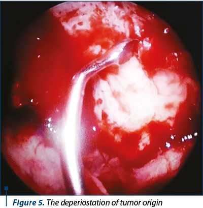
The deperiostation of the tumor attachment is required, as it lowers the risk of recurrence. We can succeed in this procedure using an angulated curette (Figure 5) or an angulated diamond burr, depending on the position of the bony stalk or hyperostosis.
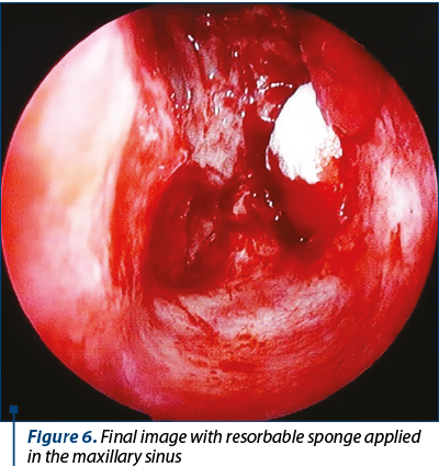
Final image of the widely opened medial maxillectomy ostium and resorbable sponge applied in the maxillary sinus. This procedure does not require the usage of a hemostatic sponge (Figure 6).
Conclusions
Inverted papilloma is a benign tumor with recurrence and malignancy potential. Recurrence is in fact a tumoral residue. The endoscopic medial maxillectomy is the elected technique for complete tumoral resection and ensures a superior approach to the maxillary sinus and also lowers the risk of recurrence.
The advantages for endoscopic medial maxillectomy include optimal exposure of maxillary and ethmoid sinuses, without the resection of any bony nasal walls, superior visualization of difficult-to-view areas, lower risk of infraorbital nerve paresthesia, lower morbidity and hospitalization, better esthetic outcomes and higher comfort for the patient, as it doesn’t require hemostatic sponges.
Conflict of interests: The authors declare no conflict of interests.
Bibliografie
- Khandekar S, Dive A, Mishra R, Upadhyaya N. Sinonasal inverted papilloma: A case report and mini review of histopathological features. Journal of Oral and Maxillofacial Pathology. 2015; 19(3); 405.
- Cunningham K, Welch K. Endoscopic medial maxillectomy. Operative Techniques in Otolaryngology. 2010; 21; 111-116.
- Sadeghi N, Al-Dhahri S, Manoukian J. Transnasal endoscopic medial maxillectomy for inverting papilloma. The Laryngoscope. 2003; 113(4); 749-753.

Asistenţa medicală dentară privată şi marketingul medical
Irina-Maria Gheorghiu, Loredana Mitran, Mihai Mitran, Alexandru Iliescu, Sânziana Scărlătescu, Paula Perlea
Asistenţa medicală dentară privată actuală se confruntă cu noţiunea de marketing. Pentru un medic nu este uşor de acceptat că practica sa privată este, din punct de vedere economic, o profesie „prestatoare de servici...
Reconstrucţia defectelor post-excizionale de pleoapă inferioară toată grosimea după chirurgia oncologică a carcinoamelor de piele
Ioana Dumitrescu, Andrada Stan, Cosmin Humulescu, Silvius Ioan Negoiță
Excizia corectă şi completă a carcinoamelor de pleoapă inferioară, cu controlul histopatologic al marginilor, duce la defecte complexe şi imprevizibile în ceea ce priveşte forma şi dimensiunea. În ace...
Tratamentul tumorilor maligne orbitare
Vladimir Sorin Ibric-Cioranu, Vlad Petrescu Seceleanu
Patologia tumorală din sfera orbitară este extrem de variată. Printre cele mai întâlnite tipuri de tumori maligne se numără carcinomul bazocelular, spinocelular şi limfomul. Procedeele de ablaţie chirurgicală cele mai folosite sunt rezecţia simplă, exenteraţia şi exenteraţia extinsă. Reconstrucţia defectelor ...
The health benefits of AudioStressNotifier: an innovative noise monitoring app and its impact on sleep
Cristina-Maria Goanţă, Ioan Burciu, Daniela Cîrpaciu, Vlad Andrei Budu
Somnul joacă un rol esenţial în menţinerea sănătăţii. Numeroşi factori pot perturba somnul, inclusiv zgomotul din mediul înconjurător. Apariţia aplicaţiilor mobile a permis dezvoltarea de solu...
Histological findings in chronic hypertrophic rhinitis
Remus Gabriel Mihalcea, Prepageran Narayanan, Erich Vyskocil, Daniela Cîrpaciu, Vlad Andrei Budu, Cristina-Maria Goanţă
Rinita cronică este o afecţiune frecventă, cu un impact semnificativ asupra calităţii vieţii. Scopul lucrării îl reprezintă examinarea modelului celular şi modificările fiziopatologice care apa...