Studiu asupra prevalenţei, diagnosticului, managementului terapeutic şi a complicaţiilor dinţilor supranumerari
Study on the prevalence, diagnosis, therapeutic management and complications of supernumerary teeth
Abstract
Aim. This descriptive study reveals clinical and paraclinical aspects related to the presence of supernumerary teeth in children with severe general disorders. Materials and method. The study based on clinical, epidemiological and radiological assessment was performed on a batch of 1651 urban patients from whom a batch of 7 patients with familial and complex history and supernumerary teeth was chosen. Results and discussion. In the study group, the most common morphological form of supernumerary tooth encountered was the mesiodens with nano crown, in the form of a stake present in four of the seven cases clinically and radiologically supervised, and the predilection was the upper jaw. The results are in line with those in the specialized literature. Conclusions. Early treatment can solve the shortcomings caused by the presence of supernumerary teeth, if the anomaly is diagnosed early. Any sign of late eruption longer than 8 months compared to the eruption of the counterpart should be investigated radiologically.Keywords
supernumerarymesiodensclinical aspectradiological aspecttreatmentRezumat
Scop. Acest studiu descriptiv relevă aspecte clinice şi paraclinice legate de prezenţa dinţilor supranumerari la copii cu afecţiuni generale grave. Materiale şi metodă. Studiul, bazat pe analiza clinică, epidemiologică şi pe evaluare radiologică, a fost efectuat într-un cabinet cu practică privată din Bucureşti, pe un lot de şapte pacienţi cu istoric familial şi general complex şi cu dinţi supranumerari. Rezultate şi discuţii. S-a constatat că meziodensul se regăseşte la ambele sexe în mod relativ egal, neexistând diferenţe majore între femei şi bărbaţi. În lotul nostru de studiu, cea mai frecventă formă morfologică de dinte supranumerar întâlnită este meziodensul cu coroană nanică, în formă de ţăruş, prezent în patru din cele şapte cazuri supravegheate clinic şi radiologic, iar localizarea predilectă este premaxilarul. Rezultatele sunt în concordanţă cu cele din literatura de specialitate. Concluzii. Tratamentul timpuriu poate rezolva neajunsurile produse de prezenţa dinţilor supranumerari, în condiţiile în care anomalia este diagnosticată devreme. Orice semn de erupţie întârziată mai mare de opt luni faţă de erupţia omologului trebuie investigat radiologic.Cuvinte Cheie
supranumerarmeziodensaspect clinicaspect radiologictratamentIntroduction
Supernumerary teeth are dental units that numerically exceed the normal dental formula, a phenomenon also known as hyperdontosis (extra teeth, duplicates, cones or aberrant teeth), which occurs in solitary or multiple forms, uniquely or bilaterally affecting both jaws.
The prevalence of supernumerary teeth ranges between 0.1% and 0.8% in temporary dentition and between 0.1% and 3.8% in permanent teeth(1-4). Boys are two times more affected than girls, and the jaw is 8.2-10 times more affected than the mandible(1-4). There is a significant association between supernumerary teeth and invaginated teeth explained by the different embryological development of the premaxillary(2).
Etiology
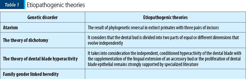
P.V. Rao(1) considers that the etiology of supernumeraries is multifactorial, involving genetic factors and environmental factors, based on theories that try to explain their occurrence (Table 1).
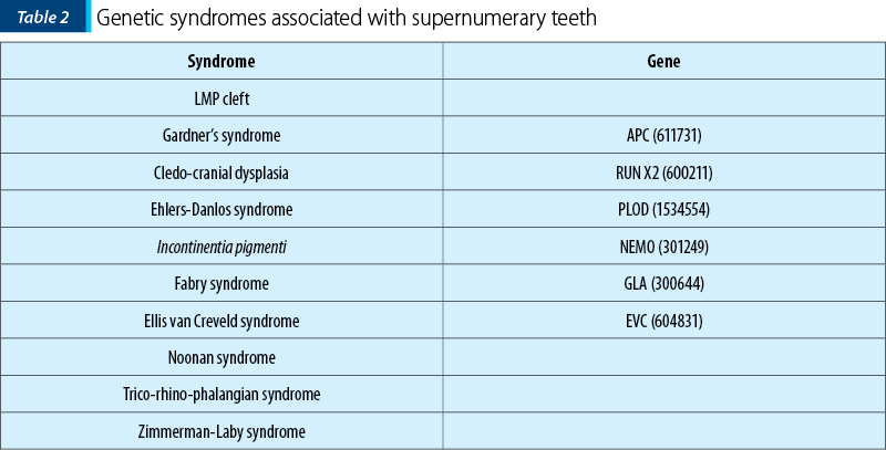
A number of genetic syndromes are associated with the presence of supernumerary teeth and the gene localization is already known(6,14) (Table 2).
Clinical and paraclinical aspects
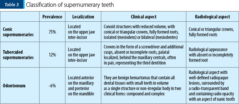
The unilateral persistence of a temporary tooth in the premaxillary area, the eruption delay, the impaction or the ectopic eruption of a permanent incisor, the severe inter-incisive diastema or the rotation of permanent incisors or dento-alveolar disharmony with frontal crowding alert the clinician to the possible presence of supernumerary teeth and require a radiological investigation to confirm the diagnosis. Their classification may be done morphologically and according to localization (Table 3)(2,3,11,8).
The most useful radiographic investigation is orthopantomography with additional images on periapical and occlusal radiographs. For the localization of a non-erupted supernumerary, parallax method is recommended with two separate X-rays from different angles for the same region or cone beam CT that provides detailed 3D imaging of the zonal structures(2,3).
Accidentally discovered in the radiological examination, the supernumerary tooth may associate complications including delayed eruption of permanent teeth in the area of interest, impaction or ectopic eruptions, severe rotations of adjacent teeth, dento-alveolar crowding or diastema which cannot be closed orthodontically, root resorption or development of follicular cysts.
Materials and method
Clinical analysis – two assessors examined the supernumerary teeth morphology through visual inspection.
Visual examination of orthopantomographies – two evaluators examined retro-dento-alveolar radiographs and orthopantomographies in the study group, with a 96.7% concordance between examiners.
Statistical analysis used the T student test and frequency analysis, which were done using SPSS 13.00 software.
The study group
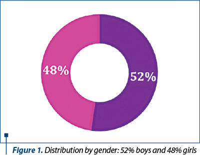
The study was performed on a group of 1651 patients, 52% boys and 48% girls, aged between 6 and 15 years old, over a one-year period (2017-2018). For the epidemiological study, the number, shape and volume of supernumerary teeth were taken into account: gender, age and ethnicity, dental history and radiological balance.
From the study group, seven cases presented mesiodens, indicating a prevalence of 0.42%. Gender distribution reveals a modest percentage in favor of male gender, but the reduced size of the affected group does not allow valid conclusions about the relatively equal affection of the two sexes.
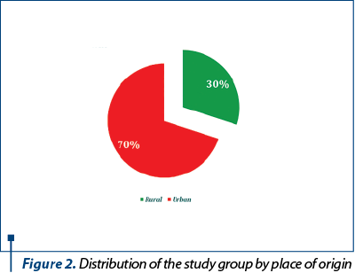
The environment of origin is dominated by the urban area with higher addressability and in the absence of dental practices in villages.
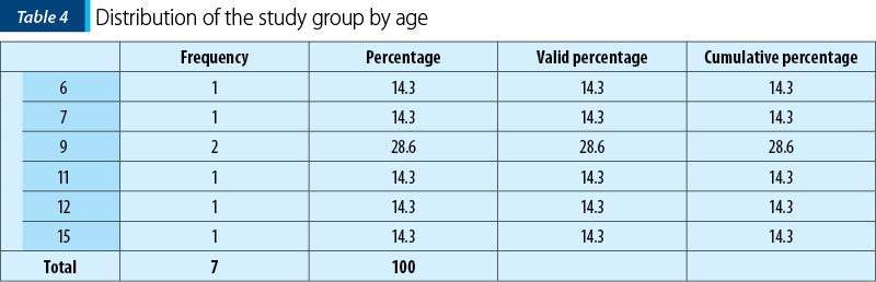
The distribution of the study group by age group is heterogeneous: the age of 9 is predominant.
Results and discussion
Although age is an important factor in assessing the prevalence of dental abnormalities for the study of supernumerary teeth, this parameter is considered to be inaccurate because there is no specific age at which supernumerary teeth begin to develop; they may occur in temporary, mixed or permanent dentition in the premaxillary region or in any region of the dental arcade. Even the absence of supernumerary teeth at a certain age does not imply, with certainty, that the patient will not present supernumeraries later; it only indicates that, at the time of the examination, there are no supernumerary dental buds(2,3).
Family history and dental history are critical factors that influence the prevalence values of supernumeraries in our study group(7,11,12,13).
Case 1
The first patient was an 11-year-old boy diagnosed with Trico-oculo-dental syndrome, the first newborn of a 38-year-old pregnant mother with type 1 diabetes mellitus developed with delayed dis-pregnancy, the birth being at term, with dystocia, by caesarean section, the newborn weighing 4100 g, with normal postnatal development.
General clinical examination reveals moderately pale skin, excessive adipose tissue on the chest and limbs, dismorphic facies, hypertelorism, epicanthus, thick eyebrows, flattened nose root, bilateral congenital cataract confirmed by olological examination, and mental retardation with QI 41 (Figure 3).
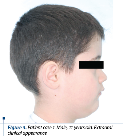
At the facial level, there is a small mouth with the corners facing downwards, a retrognathic profile. The functional examination revealed functional disorders of mastication, phonation and physiognomy.
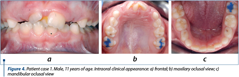
The endobucal examination (Figure 4) in mixed dentition shows, on the upper jaw median line, behind the upper central incisors, a supernumerary tooth (mesiodentes) in the form of a screwdriver, intervestibular rotations of the central incisors, a 3-mm inter-incisive diastema with divergent dental axes.
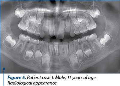
The radiological examination reveals mixed dentition, a supernumerary tooth located intraosseous, at the apex of 21, with an abnormal position, almost parallel to the occlusal plane, with the tip pointing to the crown of the dental bud of 23 (Figure 5).
Case 2
Another patient was a female aged 15 years old, the first newborn of a 35-year-old primiparous mother with a pigmented nevus in the left deltoid region, coming from an unmonitored pregnancy, the child being born on term, with a weight of 2700 g, with a good postnatal evolution. She was hospitalized in the territorial hospital at the age of 8 years old for a respiratory infectious episode. She presents skin hypopigmentation spots, concentration deficiency and psychomotor deficiency, 29 kg weight hypotrophy, papular rash in the face and upper limbs with hypochromic areas disposed along the Blaschko lines.
The dermatological examination of the skin lesions associated with the presence of neuropsychiatric disorders and corroborated with the genetic examination lead to the diagnosis of Ito hypomelanosis. Microcrania, mild facial asymmetry, and skin allergy areas are highlighted. The patient has definitive dentition, poor hygiene and a supernumerary, nanic tooth, with ectopic eruption, inter-incisive at the jaw level (Figure 6).
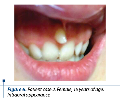
The patient was advised to have a radiological checkup to see exactly the shape and size of the root, as well as its position in relation to the surrounding tissues, and she was recommended, as treatment, the extraction of the mesiodens.
Some cases of supernumerary teeth are asymptomatic and accidentally detected at the radiological exam. Dental buds with late onset of mineralization can give a false positive diagnosis of supernumerary tooth on radiographs(7,9,11).
Case 3
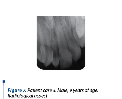
This is also the case of a 9.4-year-old male patient with marked weight hypotrophy and moderate hepatosplenomegaly, facial dismorphism, larger ears, lowered oral commissures, who was diagnosed with mucopolysaccharidosis confirmed by alpha-l iduronidase dosing in fibroblasts and leukocytes. The radiological exam revealed a supernumerary tooth included (Figure 7).
Case 4
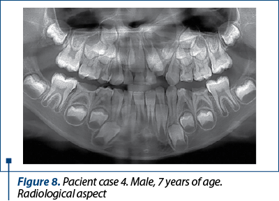
In the case of a 7-year-old male patient diagnosed with palatal cleft operated about 6 years ago and paternal heredity of congenital malformation, the orthopantomograph reveals the mesiodens located between 51 and 61, as well as the presence of a supernumerary tooth in quadrant 2. Since both 62 and the supernumerary tooth are located very close to the palatoplasty line, it was decided to send the patient to a buccal-maxillofacial surgery service (Figure 8).
The treatment of supernumerary teeth depends on the type of morphology, position, associated complications, patient’s age and dental cooperation, the radiologically appreciated radial length and the program of root-formation of the adjacent teeth, and it consists in the surgical extraction of the supernumerary tooth or, more rarely, the repositioning in the dentoalveolar arch(3,9,10).
Case 5
In a female patient, aged 12 years old, it was decided to extract the mesiodens using local anesthesia. She is the first newborn of a 30-year-old primiparous mother with minor thalassemia from a monitored pregnancy with late dis-pregnancy, the child being born on term, with a birth weight of 2400 g, with good postnatal progression. At the age of 1 year and a half, she was admitted to a pediatric hospital in Bucharest for an intercurrent superior respiratory infection; pale skin and mucosa, and splenomegaly were highlighted.
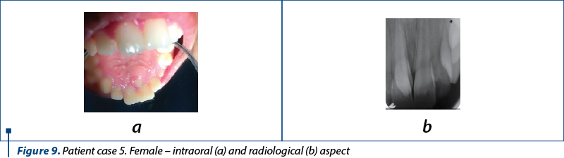
The haemogram showed hypochromic anemia, and the smear revealed an increased number of reticulocytes and cells in the form of target shooting, suggestive of thalassemia. Pathological hemoglobin electrophoresis was performed, showing a high percentage of fetal hemoglobin and A2 hemoglobin. At the age of one years old, the child returns to the pediatric-hematology department for isogroup/isoRh blood substitution-transfusion treatment. At the intraoral examination, a supernumerary tooth with a conical and sharpened appearance (Figure 9a) was observed. The patient was advised to have a radiological examination in order to assess the shape and size of the root and its position in relation to surrounding tissues (Figure 9b).
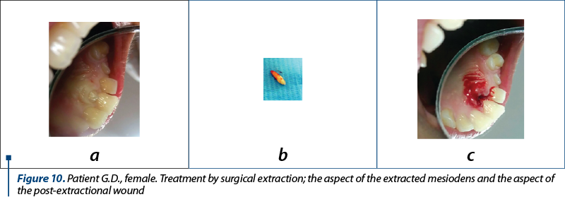
Treated through the surgical extraction of the supernumerary tooth, partially palatally erupted, with incompletely formed root, the patient was carefully monitored intrasurgically and taken care of until healing. The place of extraction was healed in about 15 days and the patient was able to resume her masticatory function without pain or discomfort (Figure 10).
In our study group, the most common morphological form of supernumerary tooth is the mesiodens with nanic crown, in the form of a stake, present in four of the seven cases clinically and radiologically monitored, as in the studies by Fadi Ata-Al and Daijit S. Gill(2,3). The number of supernumerary teeth diagnosed in two cases is double and the other cases in the study group present a single supernumerary tooth located in the jaw, as in the studies by Fadi Ata-Al and Daijit S. Gill(2,3,4,6). The predilect localization of supernumerary teeth in the batch is the maxillary inter-incisive area, as in the study by Fadi Ata-Al(2,3,6,8).
The family history and the complex, debilitating and hereditary inherited general disorders are essential variables to be reported in supernumerary teeth studies(2,3,10).
The reported prevalence of 0.42% and the equal gender distribution should be considered with caution due to the relatively small number of patients in the study group, as in the study by Anthonappa(7,11,13).
The late eruption of the maxillary incisors is associated with Caucasians, in the proportion of 20-60%, and with the presence of supernumerary teeth(2,3) especially from the tuberculed morphological type. Three of the seven cases in our study group had delayed eruptions of the adjacent upper fronts. Any sign of eruption that is more than eight months late in relation to the eruption of the counterpart should be investigated radiologically. Patients with a history of conical or tuberculed supernumeraries at younger ages have 24% chances(3) to develop unique or multiple supernumerary premolars, and this is why it is necessary to have periodic radiological monitoring in mixed teeth.
Conclusions
1. The reported prevalence of 0.42% and the equal gender distribution should be considered with caution due to the relatively small number of patients in the study group. In our study group, the most common morphological form of supernumerary tooth is the mesiodens with nanic crown, in the form of a stake.
2. The predilect localization of supernumerary teeth in the batch is the maxillary inter-incisive area.
3. Supernumerary teeth have erupted ectopically by producing dental congestion or preventing the eruption of neighbouring teeth in three of the seven cases included in the batch.
4. Supernumerary teeth produce functional disorders especially in the physiological sphere and phonation disorders.
5. Clinical and radiological examinations are essential in the diagnosis of supernumerary teeth.
6. The radiological examination identifies the presence, the coronary and root shape of the supernumerary tooth, the proximity to the permanent teeth and their stage of root formation, it allows the positive diagnosis, it orients the treatment, and it monitors the occurrence of new single or multiple supernumeraries.
7. Early diagnosis and appropriate management can minimize complications caused by supernumerary teeth.
8. The treatment indicated in patients examined is extraction and it depends on the age of the patient, dental cooperation, locally induced complications, and the root formation program of the adjacent permanent teeth.
9. Interdisciplinary management for cases in the study group is the solution for a correct diagnosis and for well-guided and periodic treatment.
10. General dentists should be informed about clinical signs and therapeutic options in the cases of supernumerary teeth.
Compliance with ethics requirements: The authors declare that all the procedures and experiments of this study respect the ethical standards of the Helsinki Declaration from 1975, as revised in 2008, as well as the national law. The informed consent was obtained from the patients included in the study.
Conflict of interests: The authors declare no conflict of interests.
Bibliografie
- Rao PV. Supernumerary molar teeth: observations in the skulls. Cent Afr J Med. 1999; 45: 324–327.
- Ata-Ali F, Ata-Ali J, Peñarrocha-Oltra D, Peñarrocha-Diago M. Prevalence, etiology, diagnosis, treatment and complications of supernumerary teeth. J Clin Exp Dent. 2014; 6(4):e414-8.
- Shah A, Gill DS, Tredwin C, Naini FB. Diagnosis and Management of Supernumerary Teeth. Dent Update. 2008; 35: 510-520.
- Gabris K, Fabian G, Kaan M, Rozsa N, Tarjan I. Prevalence of hypodontia and hyperdontia in paedodontic and orthodontic patients in Budapest. Community Dent Health. 2006; 23: 80−82.
- Guttal KS, Naikmasur VG, Bhargava P, Bathi RJ. Frequency of developmental dental anomalies in the Indian population. Eur J Dent. 2010; 4: 263–269.
- Brook AH, Elcock C, al-Sharood MH, McKeown HF, Khalaf K, Smith RN. Further studies of a model for the etiology of anomalies of tooth number and size in humans. Connect Tissue Res. 2002; 43(2-3): 289−295.
- Anthonappa RP, King NM, Rabie ABM. Diagnostic tools used to predict the prevalence of supernumerary teeth: a meta-analysis. Dentomaxillofacial Radiology. 2012; 41, 444–449.
- Khalaf K, Robinson DL, Elcock C, Smith RN, Brook AH. Toothsize in patients with supernumerary teeth and a control group measured by image analysis system. Arch Oral Biol. 2005; 50: 243−248.
- Tyrologou S, Koch G, Kurol J. Location, complications and treatment of mesiodentes − a retrospective study in children. Swed Dent J. 2005; 29: 1−9.
- Kramer PF, Feldens CA, Ferreira SH, Spiguel MH, Feldens EG. Dental anomalies and associated factors in 2- to 5-year-old Brazilian children. Int J Paediatr Dent. 2008; 18: 434–440.
- Anthonappa RP, Omer RS, King NM. Characteristics of 283 supernumerary teeth in southern Chinese children. Oral Surg Oral Med Oral Pathol Oral Radiol Endod. 2008; 105: e48–e54.
- Backman B, Wahlin YB. Variations in number and morphology of permanent teeth in 7-year-old Swedish children. Int J Paediatr Dent. 2001; 11: 11–17.
- Varela M, Arrieta P, Ventureira C. Non-syndromic concomitant hypodontia and supernumerary teeth in an orthodontic population. European Journal of Orthodontics. 2009; 31, 632–637.
- Fleming PS, Xavier GM, DiBiase AT, Cobourne MT. Revisiting the supernumerary: the epidemiological and molecular basis of extra teeth. British Dental Journal. 2010 Jan 9; 208(1):25-3.

The First National Congress of the young Romanian Society of Audiology and Communication Pathology
Sebastian Cozma
Between the 12th and 14th of September 2019, we had the joy of meeting in a special academic and cultural setting offered by the Palace of Culture in Iaşi, on the occasion of the First National Congre...
Aspecte particulare ale anatomiei etmoidale pe imagini CT
Alexandra Gheorghe, Silviu Crăc, A. Panfiloiu, Vlad Andrei Budu
Anatomia ilustrată pe descoperirile imagistice reprezintă baza înţelegerii structurilor normale şi patologice rinosinuzale. Actualmente, radiologia convenţională a fost înlocuită de imagistica CT, noul standard de au...
Utilizarea pivoturilor endodontice în restaurările odontale armate pe dinţi devitali
Irina-Maria Gheorghiu, Paula Perlea, Claudiu-Gabriel Ciolan, Loredana Mitran, Mihai Mitran, Alexandru Iliescu
În situaţiile clinice în care se impune restaurarea distrucţiilor coronare masive, este obligatorie utilizarea mijloacelor suplimentare de retenţie ce pot asigura succesul şi menţinerea restaur...
Urmărirea pacienţilor cu carcinom bazocelular
Vladimir Sorin Ibric-Cioranu, Vlad Petrescu Seceleanu
Tegumentul poate fi afectat de tumori maligne, cu preponderenţă carcinomul bazocelular, urmat de cel spinocelular şi melanomul malign. Patologia tumorală malignă din zona capului şi a gâtului se află la graniţa dintre multiple specialităţi: chirurgie maxilo-facială, dermatologie, chirurgie plasti...