Anomaliile neevolutive de structură ale smalţului dentar
Non-evolving abnormalities of enamel structure
Abstract
Purpose. Through this paper we propose the succinct presentation of the nonevolving abnormalities of the structure of enamel, from the clinical point of view, and of their etiology, as well as the awareness of the dentist and of the patients or the caregivers about the causes and complications that may occur in the case of enamel hypoplasias.Materials and method. The case of a teenager who presented in the dental office for the treatment of carious lesions based on enamel hypoplasia was studied. During the intraoral and radiological examination, the causes, the diagnosis and the appropriate treatment of the dental lesions have been established.
Results and discussion. Enamel hypoplasias are structural defects caused by the action of general or local environmental factors. They occur due to the perturbation of the apposition processes of organic matrices and their mineralization. They can affect all teeth in the case of general environmental factors, or only dental groups in case of local environmental factors. The complications are grafting of carious processes and the appearance of dental fractures.
Conclusions. Making a rigorous anamnesis accompanied by appropriate radiological examinations is essential in the case of a patient presenting hypoplasic enamel areas. It is important to distinguish hypoplasia from hypomineralization or other genetic abnormalities, pathologies that may resemble or cause similar complications.
Keywords
hypoplasiaenamelenvironmental factorscarious processRezumat
Scop. Prin această lucrare ne propunem prezentarea succintă a anomaliilor neevolutive de structură ale smalţului din punct de vedere clinic şi a etiologiei acestora, precum şi conştientizarea medicului dentist şi a pacienţilor ori aparţinătorilor privind cauzele şi complicaţiile ce pot apărea în cazul hipoplaziilor de smalţ.Materiale şi metodă. S-a studiat cazul unui pacient adolescent care s-a prezentat în cabinetul stomatologic pentru tratarea unor leziuni de tip carios pe fond de hipoplazie de smalţ. La examenul intraoral şi radiologic s-au stabilit cauzele, diagnosticul şi tratamentul corespunzător al leziunilor odontale.
Rezultate şi discuţii. Hipoplaziile de smalţ sunt defecte de structură datorate acţiunii factorilor de mediu pe cale generală sau locală. Ele apar din cauza pertubării proceselor de apoziţie a matricelor organice şi de mineralizare a acestora. Pot afecta toţi dinţii în cazul acţiunii factorilor generali de mediu sau doar unele grupe de dinţi în cazul afectării locale a factorilor de mediu. Complicaţiile sunt reprezentate de grefarea proceselor carioase şi de apariţia fracturilor dentare.
Concluzii. Efectuarea unei anamneze riguroase, însoţită de examene radiologice adecvate, este esenţială în situaţia unui pacient care prezintă zone hipoplazice de smalţ, fiind importantă diferenţierea hipoplaziei de hipomineralizare sau de alte anomalii de cauză genetică, patologii care pot semăna sau pot determina complicaţii asemănătoare.
Cuvinte Cheie
hipoplaziesmalţfactori de mediuproces cariosIntroduction
Enamel hypoplasias induced by the general action of environmental factors can manifest in both dentitions, but they occur more frequently in permanent dentition and in symmetrical, homologous teeth groups in the teeth forming the enamel during the etiological factor action(1).
The lesions are interested in certain portions of the dental surface, and not the entire dental surface, respecting the growth pattern. These are disposed parallel to the incisal edge or tip of the cusps(3).
From the clinical point of view, they are manifested by the appearance of ditches, geodes or furrows, parallel to the incisal edge or occlusal surface, which exhibit different depths and stretches in the surface depending on the duration and intensity of the etiological factor action. The dental surface level at which defects are found corresponds to the degree of formation of the enamel at the time of disruption factor action(1).
If several etiological factors occur at the time of deposition of the organic enamel matrix at different time intervals, there may be several hypoplasic type defects on the dental surface separated by the healthy dental structure. If hypoplasias are located in the vicinity of the incisional margin, there is a risk of carious graft complications or incisional edge coronary fracture(1).
The etiology is represented by factors that occur during the intrauterine life, around birth, postnatally, in the first year of life, and by allergic, infectious or chemical factors and metabolic disorders.
Prenatal rubella affects the temporary dentition, and congenital lues can cause hypoplasia-type defects because Treponema pallidum passes through the placenta and invades the fetus around the fourth month of intrauterine life, when the buds of 6-year-old molars and upper central incisors are formed. The localization of incisor defects leads to the appearance of Hutchinson teeth(1).
With regard to chemicals, tetracycline, fluoride and thalidomide are contraindicated to pregnant women because they pass through the placenta and may cause hypoplasic defects. Diabetes mellitus, endocrine disorders, maternal genital tract disorders, and the way of life of the pregnant women who frequently consume large quantities of coffee or tea can lead to the appearance of enamel structure defects(1).
Premature birth, prolonged delivery, or accidents occurring during childbirth, such as anoxia or hypoxia on the child, frequently cause structural defects that can become apparent on the neonatal line (neonatal hypoplasia). Also, the child’s various illnesses in the first year of life, such as eruptive fever, rickets, dyspepsia, low birth weight etc., can also have a trigger role(1,2,4).
The structural defects induced by the local action of environmental factors occur only at the level of one or two teeth that during the formation period came into contact with the local disturbing factor. These defects do not follow the growth pattern, not being parallel to the incisal edge or occlusal surface, but they are located at the level corresponding to the impact with the etiological factor(1).
The etiological factors are represented by temporary teeth traumas, pathology through complicated cavity of temporary teeth and other causes. Intrusion, lateral luxation and avulsion more often produce structural defects(1).
Osteitis stretched out due to the complications of dental caries reaching near to the permanent replacement tooth crypt produce structural defects in the lateral teeth in the premolar area. Other causes are due to inadequate orotracheal intubations performed on very young children with respiratory distress and inadequate radiation therapy(1).
Materials and method
The evolution of the case of a teenager who presented in the dental office for the treatment of carious lesions on the basis of enamel hypoplasia is presented. The intraoral and radiological examination established the causes, the diagnosis and the appropriate treatment of odontal lesions.
The diagnosis was established following the history combined with the direct intraoral clinical examination. The treatment was restorative, improving aesthetic and masticatory functions.
Clinical case
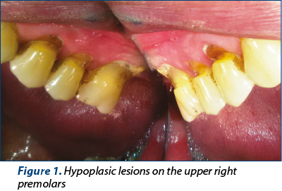

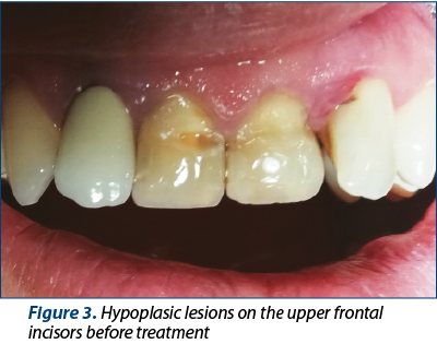
A 17-year-old male patient presented in the dentist’s office for professional brushing and for the treatment of odontal lesions. After the consultation, the presence of advanced carious lesions at the level of the teeth (Figure 1), grafted on a hypoplasic layer of enamel, has been observed. The anamnestic data revealed the poor oral hygiene of the patient (Figure 2), with the presence of yellowish-white pursy enamel areas from childhood, visible with the eruption of premolars and of inferior incisors. The personal pathological antecedents includes multiple caries in temporary teeth, respiratory infections and eruptive fevers of childhood in newborn and pre-school periods. The patient presented because of poor esthetics caused by the discoloration of the hypoplasic lesions on the upper incisors (Figure 3). We can also mention in the patient history the loss of the crown by fracture on the upper right lateral incisor that was afterwards restored by a total physionomic porcelain fused to metal crown.
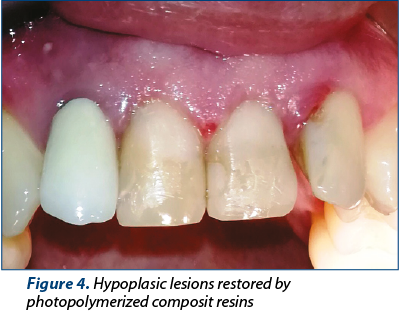
The treatment objectives consisted of periodontal stabilization by scaling, professional brushing and hygiene indications, combating dental sensitivity, dental caries prophylaxis, the rehabilitation of masticatory and aesthetic functions by restoring damaged morphological substrate, using photopolymerised composite resin materials (Figure 4).
Results and discussion
Dental enamel has a structure composed of over 90% of mineral substances and is among the toughest formations of the body. Typically, the dental enamel is thick enough to protect its substrates during a lifetime.
In the case of the action of general or local environmental factors, disturbances may occur in the normal enamel structure which, over time, cause severe complications, affecting the dentomaxillary functions: aesthetics, mastication, phonation.
Enamel hypoplasias are structural defects due to the action of the general or local environmental factors. They occur due to the perturbation of the processes of organic matrix apposition and their mineralization. They can affect all teeth in the case of the action of general environmental factors or only dental groups in case of local damage of environmental factors. The complications are grafting of carious processes and the appearance of dental fractures.
Conclusions
Making a rigorous anamnesis, accompanied by appropriate radiological examinations, is essential in the case of a patient presenting hypoplasic enamel areas. It is important to distinguish hypoplasia from hypomineralization or other genetic abnormalities, pathologies that may resemble or cause similar complications.
Compliance with ethics requirements:
The authors declare no conflict of interest regarding this article.
The authors declare that all the procedures and experiments of this study respect the ethical standards in the Helsinki Declaration of 1975, as revised in 2008, as well as the national law. The informed consent was obtained from the patient included in the study.
No funding for this study.
Bibliografie
- Luca R. Pedodonţie, vol.3. Editura Cerma. 2013; 108-125.
- Dos Santos Jr VE, et al. Early childhood caries and its relationship with perinatal, socioeconomic and nutritional risks: a cross-sectional study. BMC Oral Health. 2014; 14:47.
- Goodman AH, et al. Assessment of Systemic Physiological Perturbations from Dental Enamel Hypoplasias and Associated Histological Structures. Yearbook of Physical Anthropology. 1990; 33:59-110.
- Lai PY, et al. Enamel hypoplasia and dental caries in very-low birthweight children: a case-controlled, longitudinal study. Pediatr Dent. 1997 Jan-Feb; 19(1):42-9.

Abstracte Forum ORL.ro 2019
Lista rezumatelor lucrărilor susţinute in cadul celei de-a XI-a ediţii a Forumului ORL.ro 2019...
Aspectul endoscopic al procesului uncinat
Vlad Andrei Budu
Apofiza unciformă (procesul uncinat) reprezintă lamela anterioară a etmoidului (first lamella), fiind primul reper abordabil chirurgical endoscopic în timpul etmoidectomiei. În mod obişnuit, procesul uncinat se vizualizează prin medializarea cornetului mijlociu, dimensiunea sa permiţând ventilaţia meatului me...
Tehnici convenţionale şi moderne în reabilitarea orală a pacienţilor cu boală de reflux gastroesofagian
Cristina Bodnar, Horaţiu Bodnar, Andrei Kozma
Raportul descrie un caz clinic de boală de reflux gastroesofagian (GERD) asociat cu dentiţia uzată. Planul de tratament include metode convenţionale care utilizează şi materiale de restaurare, atât mo...
Responsabilitate și prevenție în Săptămâna Mondială a Imunizării
Florentina Ionescu
Prevenția și educația medicală continuă reprezintă fundamentul unei comunități sănătoase.
...
Bolile rare și testarea genetică, în centrul unor proiecte naționale
Cristina Ghioca
UMFCD și Institutul de Cercetare-Dezvoltare în Genomică au lansat proiectele „ExpertRARE” și „Genetică Echitabilă”. Acestea vizează, între altele, oferirea de servicii de screening genetic pentru aproape 20.000 de persoane.
...
UMF Iași a obținut acreditarea WFME pentru Medicină și Medicină Dentară
Florentina Ionescu
Acreditarea a fost obținută în urma evaluării realizate de Independent Agency for Accreditation and Rating (IAAR).
...