Din aceeași categorie
Tratamentul carcinomului renal metastatic cu terapiile TKI şi mTOR: prezentare de caz

A 66-year-old patient was admitted in the intensive care unit with neurological impairment, with 6 points on Glasgow Coma Scale. The patient had a history of chronic subdural hematoma, with recent hemorrhage and surgical intervention.
-
Automated complete blood count: severe anemia (hemoglobin 6.7 g/dl), moderate thrombocytopenia (platelet count 66,000/mm3), and leukocytosis (11,800/mm3) with monocytosis (4600/mm3).
-
Biochemistry: acute kidney injury (creatinine 7.08 mg/dl, urea 129.7 mg/dl), hyperbilirubinemia (total bilirubin 1.79 mg/dl; direct bilirubin 1.07 mg/dl), and hyperglycemia (161 mg/dl).
-
Computed tomography: multiple areas of osteolysis located in the thoracic and lumbar vertebrae, and pelvis.
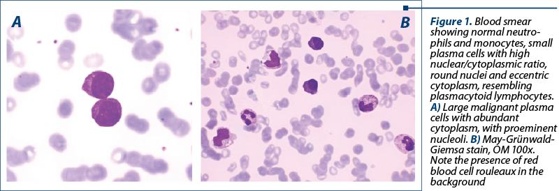
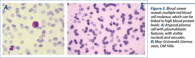
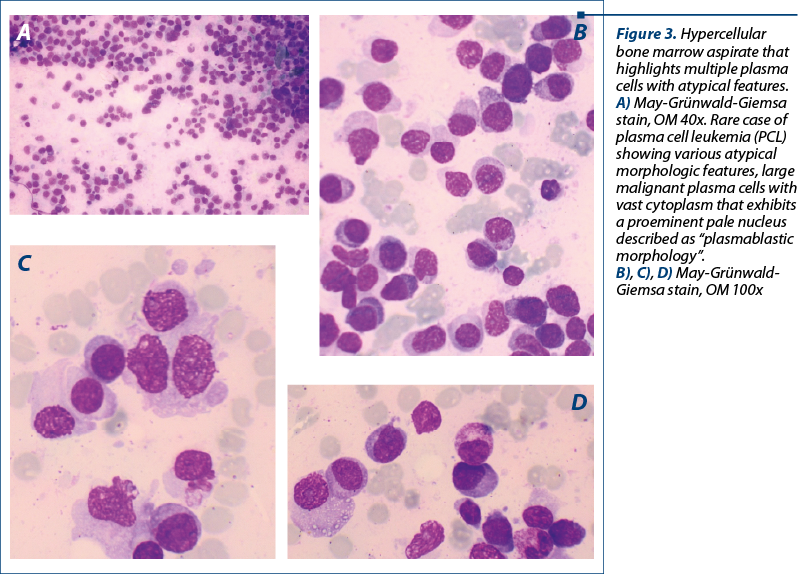
The peripheral blood smear unexpectedly revealed the presence of small-medium sized cells with high nuclear/cytoplasmic ratio, round eccentric nuclei, some with nucleoli, basophilic cytoplasm, some with vacuoles, resembling plasmacytoid lymphocytes, and also erythrocytic rouleaux, suggesting plasma cell leukemia. Bone marrow aspirate confirmed the diagnosis, describing 72% plasma cells, with plasmablastic morphology (Figures 1, 2 and 3).
Plasma cell leukemia (PCL) is a rare and aggressive variant of myeloma, characterized by the presence of circulating plasma cells. It is classified in primary or secondary PCL, the later being found in patients already diagnosed with relapsed/refractory myeloma(1).
Initially, the plasma cells are predominantly confined in the bone marrow, and only rarely enter the blood stream; the mechanisms are poorly understood. In fact, the plasma cells can be detected in the peripheral blood in only a small proportion of plasma cell dyscrasias. A major role is played by the bone marrow microenvironment(1).
According to Kyle’s criteria, the diagnosis requires circulating plasma cells (≥20% of peripheral blood leukocytes and/or an absolute count >2000/mm3 in the peripheral blood).
The diagnosis of PCL requires a thorough physical examination, along with laboratory and radiological investigations. The disease is characterized by a progressive evolution, with short survival(2-4).
leucemie cu plasmocitefrotiu de sânge perifericrulouri eritrocitare