HEMATO-ONCOLOGY
Acute myeloid leukemia cells in microscopic images
Imagini microscopice de celule leucemice mieloide
Abstract
Representative images of microscopic examination of May-Grünwald-Giemsa-stained bone marrow aspirate smears from patients with acute myeloid leukemia at diagnosisKeywords
microscopic examinationMay-Grünwald-Giemsa-stained bone marrowmyeloid leukemia cellsRezumat
Representative images of microscopic examination of May-Grünwald-Giemsa-stained bone marrow aspirate smears from patients with acute myeloid leukemia at diagnosisCuvinte Cheie
examinare microscopicăcelule leucemice mieloideMay-Grünwald-Giems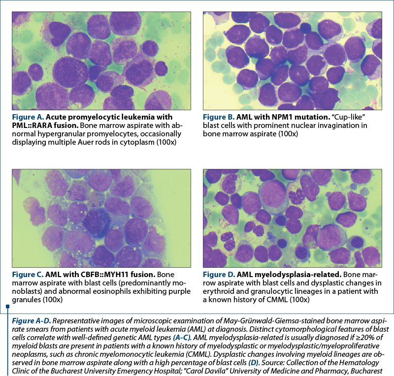
Bibliografie
Articole din ediția curentă

HEMATO-ONCOLOGY
New changes in the classification of acute myeloid leukemia proposed by WHO 2022
Silvia Badea, Emilia Severin, Diana Cîşleanu, Cristina Mambet, Claudiu Dragoş Popescu, Ana Maria Vlădăreanu
Noua revizuire a clasificării OMS (Organizaţia Mondială a Sănătăţii) din 2022 a leucemiilor acute mieloide (LAM) are ca scop evidenţierea noilor date obţinute în ultimii ani cu privire la patogeneza b...
REVIEW
Some important trials regarding the digestive cancers presented at ASCO and ESMO congresses with impact in current medical practice
Alexandru C. Grigorescu
Acest scurt review se referă la principalele studii prezentate la congresele ASCO şi ESMO din 2023, având ca subiect cancerele digestive. Din analiza acestor studii rezultă că chimioterapia, în specia...
REVIEW
Analysis of abstracts sent to the 2023 Congress of the National Society of Medical Oncology
Alexandru C. Grigorescu
Acest review include o analiză sumară a rezumatelor trimise la Congresul Societăţii Naţionale de Oncologie Medicală din anul 2023. Astfel, din cele 49 de rezumate trimise, 26 au fost prezentări de caz, 12 au fost review-...Articole din edițiile anterioare
HEMATO-ONCOLOGY
New changes in the classification of acute myeloid leukemia proposed by WHO 2022
Silvia Badea, Emilia Severin, Diana Cîşleanu, Cristina Mambet, Claudiu Dragoş Popescu, Ana Maria Vlădăreanu
Noua revizuire a clasificării OMS (Organizaţia Mondială a Sănătăţii) din 2022 a leucemiilor acute mieloide (LAM) are ca scop evidenţierea noilor date obţinute în ultimii ani cu privire la patogeneza b...
CASE PRESENTATION
Refractory immune thrombocytopenia – case presentation
Iuliana Iordan, Andreea Neculcea, Stejara Nicoleta Mihai, Diana Emanuela Bonea, Andreea Spînu, Alina Mititelu, Claudiu Popescu, Raluca Truican, Anca Nicolescu, Ana Maria Vlădăreanu
Trombocitopenia imună este o boală autoimună care implică distrugerea trombocitelor în splină. Adesea, pacienţii nu răspund la ter...
HEMATO-ONCOLOGY