Adult T-cell leukemia/lymphoma (ATLL) is an aggressive neoplasm of the T-cells caused by the infection with human T-cell lymphotropic virus type-1 (HTLV-1). ATLL is classified into four subtypes, namely acute, lymphoma, chronic, and smoldering. The acute, lymphoma and chronic unfavorable types are considered aggressive forms of ATLL. We present some peripheral blood smear images of a patient diagnosed with acute type ATLL.
HEMATO-ONCOLOGY
Images in hematology: adult T-cell leukemia/lymphoma
Imagini în hematologie: leucemia/limfomul cu celule T al adultului
First published: 27 martie 2024
Editorial Group: MEDICHUB MEDIA
DOI: 10.26416/OnHe.66.1.2024.9402
Abstract
Rezumat
Leucemia/limfomul cu celule T al adultului (ATLL) este un neoplasm agresiv al celulelor T, cauzat de infecţia cu virusul limfotrop de tip 1 al celulelor T umane (HTLV-1). ATLL este clasificat în patru subtipuri, şi anume: acut, limfom, cronic şi smoldering. Tipurile acut, limfom şi cronic nefavorabil sunt considerate forme agresive de ATLL. În acest articol, prezentăm imagini cu frotiul de sânge periferic al unei paciente diagnosticate cu ATLL forma acută.
A 72-year-old female patient was brought to the Emergency Room of the Emergency University Hospital Bucharest, Romania, for altered clinical and mental status. She had a medical history of Alzheimer’s disease and hypertension. The clinical and laboratory data revealed laterocervical adenopathy, splenomegaly, leukocytosis with lymphocytosis, mild thrombocytopenia, acute kidney injury, and hypercalcemia. She was admitted to the hematology clinic for diagnosis and treatment.
Laboratory findings
Complete blood count: high white blood cell count (27,000/mm3), hemoglobin within normal range (12.7 g/dl), mild thrombocytopenia (114,000/mm3).
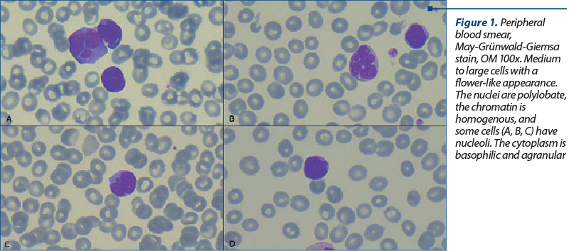
Peripheral blood smear: 35% atypical lymphocytes (Figure 1).
Biochemistry studies: normal liver function, hyperuricemia (uric acid 14.7 mg/dl), creatinine 2.59 mg/dl, hypercalcemia 19.48 mg/dl, lactate dehydrogenase level was twofold higher than the normal range.
Anti-HTLV-1 antibodies – positive.
The computed tomography scan showed multiple adenopathies, with the largest diameter of 4.4 cm, and hepatosplenomegaly.
Immunophenotyping by flow cytometry was performed. It revealed the presence of approximately 40% atypical T-cells: low expression of CD3 and absence of CD7, CD4, CD25, and CD45RO. The results confirmed the diagnosis of acute-type adult T-cell leukemia/lymphoma.
Adult T-cell leukemia/lymphoma (ATLL) is an aggressive hematologic neoplasm of T-cells caused by infection with the human T-cell lymphotropic virus(1). According to Shimoyama criteria, ATLL is classified into four subtypes – namely, acute, lymphoma, chronic, and smoldering(2).
The laboratory diagnosis of ATLL is based on clinical, morphological and immunophenotypic characteristics, besides confirming the HTLV-1 infection(3). The morphological exam often describes a heterogeneous population of lymphocytes. The atypical lymphocytes have variable sizes, from small to large, irregularly contoured, and hyperlobulated nucleus, giving the cell a flower-like appearance, with prominent nucleoli and basophilic cytoplasm, sometimes with vacuoles(4).
The acute subtype of ATLL is the most common one, occurring in 60% of cases(3). It is characterized by leukemic manifestations, tumoral lesions, and 1% or more atypical lymphocytes in the peripheral blood(2). The survival is short, usually less than one year, despite aggressive treatment(3).
Corresponding author: Ana-Maria Vlădăreanu E-mail: vladareanuanamaria@yahoo.com
Conflict of interest: none declared.
Financial support: none declared.
This work is permanently accessible online free of charge and published under the CC-BY licence.

Bibliografie
-
Iwanaga M, Watanabe T, Yamaguchi K. Adult T-cell leukemia: a review of epidemiological evidence. Front Microbiol. 2012;3:322.
-
Shimoyama M. Diagnostic criteria and classification of clinical subtypes of adult T-cell leukaemia-lymphoma. Br J Haematol. 1991;79(3):428-437.
-
Freedman AS, Aster JC, Suzuki R. Clinical Manifestations, Pathologic Features, and Diagnosis of Adult T Cell Leukemia-Lymphoma. UpToDate. 2021. www.uptodate.com
-
Dahmoush L, Hijazi Y, Barnes E, Stetler-Stevenson M, Abati A. Adult T-cell leukemia/lymphoma: A cytopathologic, immunocytochemical, and flow cytometric study. Cancer. 2002;96(2):110-116.