Imagini în hematologie: leucemia cu plasmocite
Images in hematology: plasma cell leukemia
Abstract
We present the images of peripheral blood smears of a patient diagnosed with plasma cell leukemia. Plasma cell leukemia is a rare and aggressive disease, characterized by more than 20% plasma cells in the peripheral blood or/and an absolute count of more than 2000/mm3.Keywords
plasma cell leukemiaperipheral blood smearerythrocytic rouleauxRezumat
Prezentăm imaginile cu frotiurile de sânge periferic ale unui pacient diagnosticat cu leucemie cu plasmocite. Leucemia cu plasmocite este o boală rară şi agresivă, caracterizată prin prezenţa a mai mult de 20% de plasmocite în sângele periferic şi/sau peste 2000/mm3 în valoare absolută.Cuvinte Cheie
leucemie cu plasmocitefrotiu de sânge perifericrulouri eritrocitareA 66-year-old patient was admitted in the intensive care unit with neurological impairment, with 6 points on Glasgow Coma Scale. The patient had a history of chronic subdural hematoma, with recent hemorrhage and surgical intervention.
-
Automated complete blood count: severe anemia (hemoglobin 6.7 g/dl), moderate thrombocytopenia (platelet count 66,000/mm3), and leukocytosis (11,800/mm3) with monocytosis (4600/mm3).
-
Biochemistry: acute kidney injury (creatinine 7.08 mg/dl, urea 129.7 mg/dl), hyperbilirubinemia (total bilirubin 1.79 mg/dl; direct bilirubin 1.07 mg/dl), and hyperglycemia (161 mg/dl).
-
Computed tomography: multiple areas of osteolysis located in the thoracic and lumbar vertebrae, and pelvis.
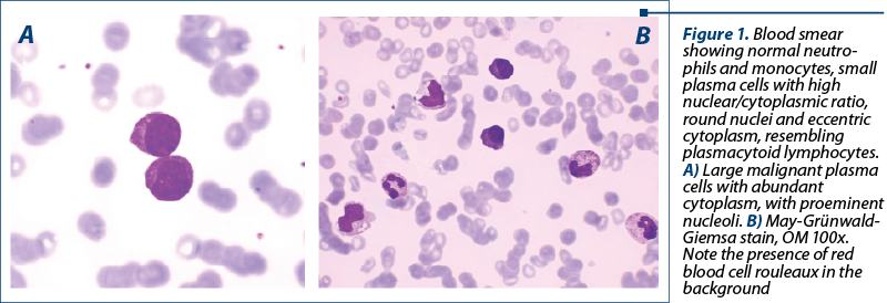
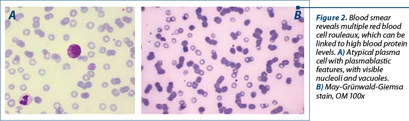
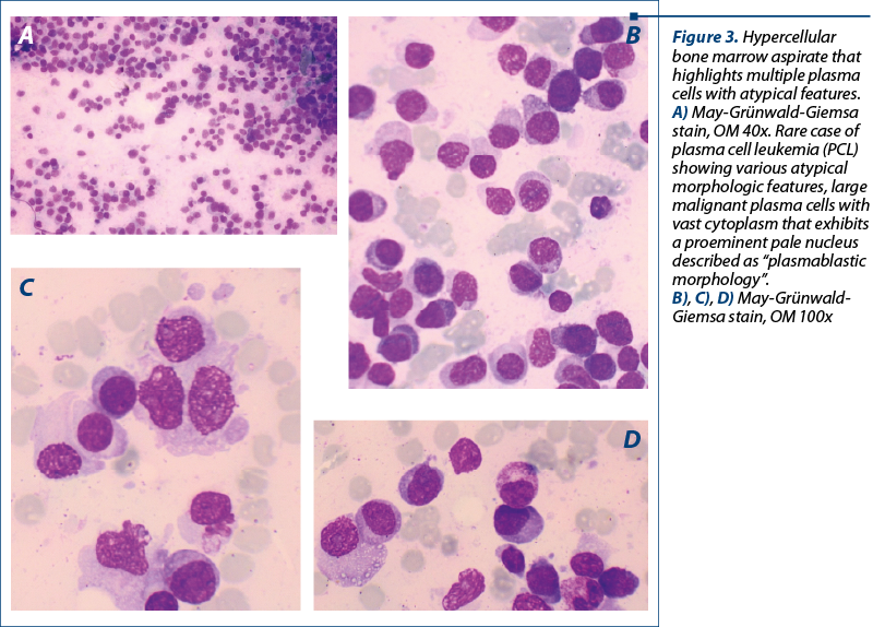
The peripheral blood smear unexpectedly revealed the presence of small-medium sized cells with high nuclear/cytoplasmic ratio, round eccentric nuclei, some with nucleoli, basophilic cytoplasm, some with vacuoles, resembling plasmacytoid lymphocytes, and also erythrocytic rouleaux, suggesting plasma cell leukemia. Bone marrow aspirate confirmed the diagnosis, describing 72% plasma cells, with plasmablastic morphology (Figures 1, 2 and 3).
Plasma cell leukemia (PCL) is a rare and aggressive variant of myeloma, characterized by the presence of circulating plasma cells. It is classified in primary or secondary PCL, the later being found in patients already diagnosed with relapsed/refractory myeloma(1).
Initially, the plasma cells are predominantly confined in the bone marrow, and only rarely enter the blood stream; the mechanisms are poorly understood. In fact, the plasma cells can be detected in the peripheral blood in only a small proportion of plasma cell dyscrasias. A major role is played by the bone marrow microenvironment(1).
According to Kyle’s criteria, the diagnosis requires circulating plasma cells (≥20% of peripheral blood leukocytes and/or an absolute count >2000/mm3 in the peripheral blood).
The diagnosis of PCL requires a thorough physical examination, along with laboratory and radiological investigations. The disease is characterized by a progressive evolution, with short survival(2-4).
Bibliografie
-
Rich R, Fleisher T, Shearer W, Schroeder H, Frew A, Weyard C. Monoclonal Gammopathies, Clinical Immunology: Principles and Practice. Fifth Edition, Elsevier, 2019. doi: 10.1016/B978-0-7020-6896-6.00108-3.
-
Ravi P, Kumar SK, Roeker L, Gonsalves W, Buadi F, Lacy MQ, Go RS, et al. Revised diagnostic criteria for plasma cell leukemia: results of a Mayo Clinic study with comparison of outcomes to multiple myeloma. Blood Cancer J. 2018;8(12):116. doi: 10.1038/s41408-018-0140-1.
-
Fernández de Larrea C, Kyle RA, Durie BG, Ludwig H, Usmani S, Vesole DH, et al. International Myeloma Working Group. Plasma cell leukemia: consensus statement on diagnostic requirements, response criteria and treatment recommendations by the International Myeloma Working Group. Leukemia. 2013;27(4):780-791. doi: 10.1038/leu.2012.336.
-
Vlădăreanu AM. Diagnosticul hemopatiilor maligne. In: Note şi imagini de atlas. Editura Universitară „Carol Davila”, Bucureşti, 2007.

Abstracts for IOB Scientific Days
...
Limfom non-Hodgkin cu evoluţie agresivă – prezentare de caz
Minodora Onisâi, Iuliana Iordan, Ana Maria Neagu, Iuliana-Maria Nicorescu, Ana Maria Vlădăreanu
Limfomul folicular este cel mai frecvent limfom non-Hodgkin indolent....
Corelaţii între biomarkerii cancerului colorectal
Alin Radu, Alexandru Grigorescu
Acest scurt review descrie principalii biomarkeri utilizaţi în cercetare şi în practica clinică pentru cancerul de colon....
Acute myeloid leukemia cells in microscopic images
Stejara Nicoleta Mihai, Cristina Enache, Cristina Mambet, Ana Maria Vlădăreanu
Representative images of microscopic examination of May-Grünwald-Giemsa-stained bone marrow aspirate smears from patients with acute myeloid leukemia at diagnosis...
Refractory immune thrombocytopenia – case presentation
Iuliana Iordan, Andreea Neculcea, Stejara Nicoleta Mihai, Diana Emanuela Bonea, Andreea Spînu, Alina Mititelu, Claudiu Popescu, Raluca Truican, Anca Nicolescu, Ana Maria Vlădăreanu
Trombocitopenia imună este o boală autoimună care implică distrugerea trombocitelor în splină. Adesea, pacienţii nu răspund la ter...