Computed-tomography study of the ethmoid labyrinth. A study on 192 ethmoid lateral masses
Studiu computer-tomografic al labirintului etmoidal. Un studiu pe 192 de mase laterale etmoidale
Abstract
The ethmoid sinuses are pneumatic cavities situated in the lateral masses of the ethmoid bone. High variability of ethmoid development, often creates confusion and uncertainty among sinus surgeons. The objective of this study was to measure different important ethmoid mass dimensions using computed-tomography on 192 ethmoid lateral masses. We measured the anterior-posterior dimensions of the ethmoid lateral mass and of the anterior ethmoid, and also the height of the ethmoid mass in relation to the lamina cribrosa and skull base. No significant differences were observed between dimensions on the right and left sides.Keywords
ethmoid sinusdimensionsvariabilitycomputed-tomographyRezumat
Sinusurile etmoidale sunt cavităţi pneumatice situate în grosimea maselor laterale ale etmoidului. Variabilitatea mare de pneumatizare etmoidală poate să creeze adeseori confuzii şi neclarităţi între chirurgiii care practică chirurgie rinosinusală. Obiectivul studiului a fost să măsoare diferitele dimensiuni importante la 192 de mase laterale ale etmoidului. Au fost măsurate dimensiunile antero-posterioare ale maselor laterale etmoidale şi ale etmoidului anterior, cât şi înălţimea masei laterale a etmoidului în raport cu lama ciuruită şi tavanul etmoidal. Nu au fost observate diferenţe semnificative între părţile dreaptă şi stângă.Cuvinte Cheie
sinus etmoidaldimensiunivariabilitatetomografie computerizatăIntroduction
The ethmoid sinuses are pneumatic cavities situated inside the lateral masses of the ethmoid bone. The ethmoid sinuses consist of air cells structured like a honeycomb inside the lateral masses of the ethmoid bone, sometimes developing outside its bony limits. In the 5th and 6th months of intrauterine development and extension of the nasal epithelium in the lateral nasal wall takes place, which will determine the apparition of ethmoid air cells. After this moment cells begin to develop and enlarge, being pneumatized through their origin. This will also be the ostium of each cell. This is considered to be the primary pneumatization of the ethmoid cells(1,2,3,4).
Pneumatization of ethmoidal cells is highly controversial. Most modern authors consider that it basically consists of two phases, the primary and secondary types, when cells initially appear as invaginations on the lateral nasal wall and then start to develop and grow. The growth pattern of these cells is not specific and varies among individuals.
Knowledge about the anatomy of the ethmoid is crucial for endoscopic sinus surgeons, because the ethmoid is currently a surgical pathway to all other paranasal sinuses (1,4,5,6,7).
Materials and methods
The authors present a descriptive study developed in our department. We examined a total of 96 preserved adult human skulls (n=96) from the collection of Craniology of the “Fr.I.Rainer” Institute for Anthropology in Bucharest. The skulls were chosen from the adult skull collection and were specifically distributed in two equal groups for male and female, so we had 48 female and 48 male skulls.
The skulls were scanned with high-resolution computed-tomography, using a protocol with 0.6 mm slices for the paranasal sinus region. The data were afterwards imported in OsiriX software, where three-dimensional reconstructions were performed. We measured the dimensions of the ethmoid sinuses on both sides in all axial, sagittal and frontal planes. The measurements included the dimensions of the anterior ethmoid and also of the whole ethmoid mass, considering the limit between the two, the basal lamella of the middle turbinate.
Results
From the 96 preserved human skulls used for examination, 48 were male and 48 female. The age of the skulls varied between 30 to 60 years of age, with an average of 44.2 years.
The data we received after examination showed a maximum anterior to posterior length of the ethmoid lateral mass of 4.62 cm as measured in the transverse plane and 4.53 cm in the sagittal plane (Figures 1, 2). The minimum anterior to posterior dimension of the ethmoid lateral mass was also available and showed values of 2.96 cm in the transverse plane and 2.87 cm in the sagittal plane, respectively. Average anterior to posterior dimension of the ethmoid lateral mass was calculated and revealed a value of 3.80 cm in the transverse plane and 4.007 cm in the sagittal plane.
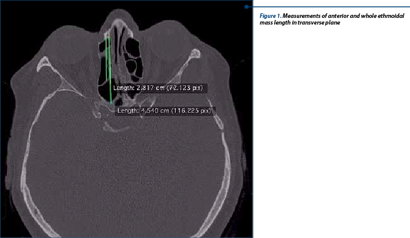
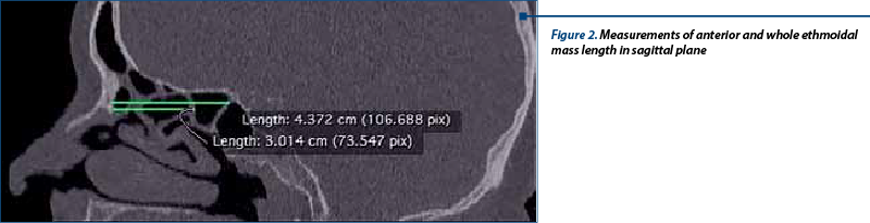
The anterior to posterior dimensions of the anterior ethmoid measured to the level of the basal lamella of the middle turbinate were also evaluated and showed values as follows: the minimum dimension was 2.034 cm in the transverse plane and 2.064 cm measured in the sagittal plane. The maximum dimension was 3.57 cm in the transverse plane and 3.41 cm in the sagittal plane. Average anterior to posterior dimension of the anterior ethmoid was 2.68 cm measured in the transverse plane and 2.77 cm in the sagittal plane.
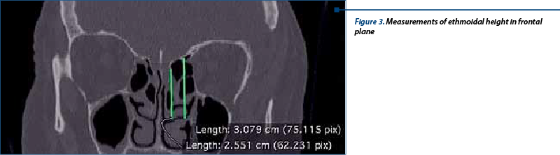
The height of the ethmoid lateral mass was measured in two different planes, according to the ethmoid roof and the lamina cribrosa (Figure 3). The inferior limit of the ethmoid mass was considered to be the inferior aspect of the middle turbinate. According to our study, the height of the ethmoid mass, measured to the roof of the ethmoid showed minimum and maximum values of 2.57 cm and 3.214 cm respectively. Average height of the ethmoid sinus measured in this plane was 2.98 cm. Measuring the height of the ethmoid sinus to the level of the lamina cribrosa, different measurements occurred: the minimum and maximum heights were 2.55 cm and 3.34 cm respectively. Average height of the ethmoid in this plane was 2.94 cm.
Discussions
Computed-tomography is the gold standard in evaluating the arrangement and dimensions of the ethmoid sinuses, as for all paranasal sinuses. Ethmoid lateral mass dimensions are important information in functional endoscopic sinus surgery due to the great variability of ethmoid extension and pneumatization. Computed-tomography started to be used in evaluation of paranasal sinuses since the early 1990’s and it has become mandatory in the pre-operative stage(8,9,10).
To our knowledge, there are no statistical reports about the exact dimensions of the ethmoid labyrinth. In one study, Rozylo-Kalinowska et al., described three types of ethmoid pneumatization, in relation to the lacrimal sac. According to their study, the most common morphology of the ethmoid sinus was the second type, when ethmoid cells extend anteriorly to the posterior lacrimal crest, but remain posterior to the fronto-lacrimal suture(11).
Measurements of the ethmoid sinuses have proved to be difficult using computed-tomography due to subjective assessment of the operator, and the differences of the same measurement in two planes of section (transversal and/or sagittal). In our study, the differences were of minimal value. We consider that standardized planes of section should be implemented in order to be accurate in these measurements.
Conclusions
Computed-tomography is the preferred method for examining the morphology and dimensions of the paranasal sinuses, due to the significant detail and resolution it offers.
Nowadays, computed tomography is also practically a reachable and inexpensive technique. It can be used to measure different dimensions of the paranasal sinuses, in all three planes of section.
The anterior-posterior dimensions of the ethmoid lateral mass vary according to our study between 2.96 cm and 4.62 cm. The anterior-posterior dimensions of the anterior ethmoid (considering the basal lamella of the middle turbinate as the limit between the anterior and posterior ethmoidal cells) are between 2.034 cm and 3.57 cm.
The height of the ethmoid lateral mass was also evaluated in the study and 2.57 cm and 3.214 cm. No significant differences were observed between right and left sides.
Acknowledgement: This paper is supported by the Sectorial Operational Program Human Resources Development (SOP HRD), financed from the European Social Fund and by the Romanian Government under the contract number POSDRU/159/1.5/S/137390/.
Bibliografie
-
H.R.Stammberger, D.W.Kennedy. Paranasal sinuses :anatomic terminology and nomenclature. Annals of Otology,Rhinology and Laryngology Suppl. 167, vol104, no 10, Part 2, pp7-16 -1995
-
Ozum Tuncyurek, Hulya Eyigor, Murat Songu. The relationship among concha bullosa, septal deviation and chronic rhinosinusitis. J Med Updates 2013:3 (1) :1-7
-
W.E Bolger, C.A Butzin, D.S Parsons. Paranasal sinus bony anatomic variations and mucosal abnormalities: CT analysis for endoscopic sinus surgery. Laryngoscope 1991. 101:56-64
-
S.J Zinreich, D.E Mattox, D.W. Kennedy, et al. Concha bullosa :CT evaluation. J Comput Assist Tomogr 1988; 12:778-84
-
H.H Unlu, S Akyar, R.Caylan, Y. Nalca. Concha bullosa. J Otolaryngol 1994; 23:23-7
-
Hatice Gul Hatipoglu, Mehmet Ali Cetin, Enis Yuksel. Concha bullosa types: their relationship with sinusitis, ostiomeatal and frontal recess disease. Diagn Intervent Radiol 2005; 11:145-149
-
M. Lidov, P.M. Som. Inflammatory disease involving a concha bullosa (enlarged pneumatized middle nasal turbinate):MR and CT appearance. AJNR Am J Neuroradiol 1990; 11:999-1001
-
Sahlstrand-Johnson et al.: Computed tomography measurements of different dimensions of maxillary and frontal sinuses. BMC Medical Imaging 2011 11:8.
-
White PS, Robinson JM, Stewart IA, Doyle T: Computerized tomography mini-series: an alternative to standard paranasal sinus radiographs. Aust N Z J Surg 1990, 60(1):25-29.
-
Christiana Maia Nobre Rocha de Miranda, Carol Pontes de Miranda Maranhão, Fabiana Maia Nobre Rocha Arraes, Igor Gomes Padilha, Lucas de Pádua Gomes de Farias, Mayara Stephanie de Araujo Jatob, Anna Carolina Mendonça de Andrade, Bruno Gomes Padilha - Anatomical variations of paranasal sinuses at multislice computed tomography: what to look for. Radiol Bras 2011, 44(4).
-
Rozylo-Kalinowska I, Jedrezejewski G, Burdan F. Types of ethmoid sinus morphology on the basis of computed-tomography examination. Folia Morphol 2003, 62(4):407-409.

Abstracte Forum ORL.ro 2015
Lista rezumatelor lucrărilor susţinute in cadul celei de-a V-a ediţii a Forumului ORL.ro 2015...
Cancerul foselor nazale şi al sinusurilor paranazale - Caz clinic: formaţiune tumorală nazoetmoidală dreaptă - carcinom adenoid chistic slab diferenţiat
Bogdan Mocanu, Vlad Andrei Budu, Irina Boagiu, Denisse Grigorescu, Silviu Oprescu, A. Coman, Daniel Mirea
Pacienta C.E., în vârstă de 68 de ani, se internează pentru obstrucţie nazală persistentă predominant dreapta, apărută de aproxima...
Paroxismia vestibulară - compresia vasculonervoasă a nervului cohleo-vestibular
Mădălina Georgescu
Paroxismia vestibulară reprezintă una din etiologiile sindromului vestibular periferic, manifestat prin crize repetitive de vertij cu durată de câteva minute şi tinitus. ...
Endoscopic approach of rinosinusal mucocele
Vlad Andrei Budu, I. Bulescu, Tatiana Decuseară, Diana Cojocaru, Alexandra Schnaider, Bogdan Mocanu
Autorii au realizat un studiu retrospectiv bazat pe experienţa clinică acumulată prin urmărirea a 93 de pacienţi internaţi în IFACF-ORL „Prof. Dr. D. Hociota” - Secţia I-IIA, între ianuarie 2010 şi ianuarie 2015 pentru m...
Sfaturi şi trucuri în etmoidectomia endoscopică
Vlad Andrei Budu, Alexandra Schnaider, Bogdan Mocanu, I. Bulescu
Etmoidectomia endoscopică este tehnica chirurgicală care constă în deschiderea întregului labirint etmoidal pentru a asigura drenajul favorabil al sinusurilor paranazale. Sunt câteva tehnici de realizare a etmoidectomiei...