Din aceeași categorie

Opinii şi recomandări curente pentru utilizarea meşei în chirurgia ginecologică – review al literaturii
1. Introduction
Uterine artery embolization (UAE), or uterine fibroid embolization (EFM), a technique introduced in the treatment of uterine fibroids in the 1990s, has been increasingly used in recent decades. The results of several trials appeared in the middle of the 2000-2010 decade, including those of Mara et al. (2006) randomized controlled trials, the REST and EMMY trials(1-4).
All of these studies reported an improvement of symptoms and of the quality of life following UAE, but they also reported a 28-32% reintervention rate after this initial procedure(3,4). However, for many women of reproductive age, seeking for conservative fibroid management, UAE has become a valid option, which led to an yearly increase in the number of this type of procedure. The accumulation of data on the results of this procedure revealed, besides the expected benefits, also side effects and complications, initially unanticipated(5,6). In order to provide a proper management of the uterine fibroid, both patient and physician should be aware and informed about all UAE possible effects. In fact, all treatment methods for uterine fibroids have advantages and disadvantages and, in order to help the physician and the patient to decide, national or even international dedicated registries should be created for comparing data on different treatment options for uterine fibroids(7,8).
The aim of this study was to evaluate the complications and side effects due to the foreign material on the myometrium and ovaries and, thus, the unwanted consequence of UAE, starting from a series of three cases.
2. Presentation of the case series
2.1. Case 1
A 41-year-old woman was admitted in our unit (“Sf. Ioan” Emergency Clinical Hospital, Bucur Maternity, Bucharest) for severe pelvic-abdominal pain. The symptoms started many years before and progressively increased in intensity in the last previous days. From the medical documents, we noticed that, eight years before, the patient underwent myomectomy for uterine fibroids, followed by UAE four years ago, for the reappearance of multiple fibroids.
The medical report indicated that polyvinyl alcohol (PVA) particles were used for UAE. The physical gynecological examination revealed globally enlarged uterus, similar to a 12-week pregnancy size, irregular, difficult to delimit, flanked to the left border by a structure of cystic consistency of approximately 9/8 cm. Abdominal and transvaginal ultrasound scan indicated enlarged uterus (97/75/50 mm), irregular, in an intermediate position, myometrium with inhomogeneous structure, and irregular thin endometrium; on the left board of the uterus there was a 87/79 mm structure with mixed appearance, both solid and liquid, without Doppler signal acoustic shadow and that could not be mobilized, suggesting adherence to the uterus.
At the general physical examination, there were noted grade III obesity (BMI 35.6) and varicose veins of the lower limbs. The surgical treatment was chosen, and at the surgery, an adhesion syndrome with the pelvis blocked by adhesions between the omentum, ileum, sigmoid colon, rectum and genitals was found. After lysis of adhesions, there could be noticed: an enlarged and irregular uterus, one endometriotic cyst in the left ovary of approximately 9 cm in diameter, multiple foci of endometriosis in the ovary and right salpinges, as well as disseminated in the pelvic area. Total hysterectomy with bilateral anexectomy was performed. The evolution was favorable and the patient was discharged on the seventh day postoperatively.
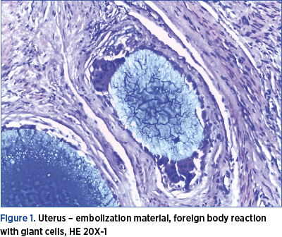
In addition to confirming endometriosis and adenomyosis, the histopathological exam revealed “diffuse fibrosis in the wall thickness, vascular structures with thickened wall and intraluminal amorphous material, perivascular inflammatory infiltration with giant multinucleate histiocytes of foreign body. Histological appearance of nonspecific giant-cell granulomatous inflammation” (Figure 1).
2.2. Case 2
A 52-year-old patient came to the “Sf. Ioan” Emergency Clinical Hospital, Bucur Maternity, Bucharest, for pelvic pain and irregular bleeding not responding to treatment. From the anamnesis, it was recorded that about 3 years ago she had been treated by a uterine artery embolization session, with polyvinyl alcohol particles, for multiple uterine fibroids. The gynecological and ultrasound examination confirmed the existence of an enlarged uterus, of 16/14/10 cm in volume, by the presence of several formations with dimensions up to 5 cm in diameter. The patient opted for total hysterectomy with bilateral adnexectomy. Pelvic adhesion syndrome was found intraoperatively. The postoperative evolution was good, the patient being discharged on the sixth postoperative day.
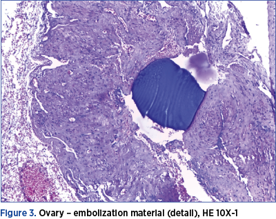
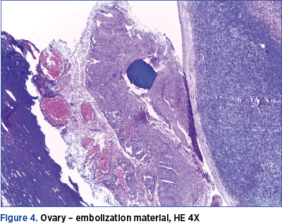
At the histopathological examination, beside the confirmation of the presence of fibroids, there were found vascular intraluminal amorphous material, vascular and perivascular infiltration with giant multinucleated histiocytes, diffuse fibrosis in the thickness of the uterine wall and also intravascular foreign body in the ovaries, with inflammatory reaction of the same type (Figures 2, 3 and 4).

2.3. Case 3
A 29-year-old patient presented herself at the “Sf. Ioan” Emergency Clinical Hospital, Bucur Maternity, Bucharest, for a uterine tumor, that was supposed to be a uterine fibroid of approximately 9 cm in diameter.
Following the anamnesis, it was recorded that, four years before, she had an UAE for a fibroid of about 8 cm in diameter and then, one year ago, she repeated the procedure because, after an initial reduction in size, to 5-6 cm in diameter, the uterine tumor began to grow again.
At her first UAE, there were used 700-900-micron gelatin microspheres (Embosphere®) and fragments of gelaspon for the second PVA particles. However, even after the second UAE, the tumor continued to grow. The patient was administered GnRH analogues for six months, but without remarkable results, and after that she was operated and myometrectomy was performed.
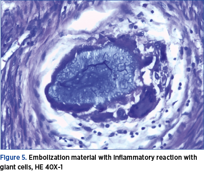
Intraoperatively, the tumor removal was difficult because it had an uneven, soft consistency and the uterine wall was also altered in terms of consistency and it was not possible to find a satisfactory cleavage space. Postoperatively, the evolution was favorable, the patient being discharged on the sixth day. The histopathological examination confirmed the diagnosis of uterine leiomyoma, but there were also observed in the surrounding myometrium the thickening of vascular structures containing intraluminal amorphous material, with vascular and perivascular infiltration with giant multinucleated histiocytes. The histological aspect corresponds to nonspecific granulomatous inflammation with giant cells (Figure 5).
3. Discussion
Historically, it is cited that the first successful embolization of the uterine arteries, for persistent metrorrhagia, in a woman with uterine fibroids, was performed in 1974, in Paris, by Dr. Jean-Jaques Merland(9). Afterwards, this method was adopted by Dr. Jacques Ravina, who initially used it preoperatively, to improve the conditions for performing myomectomy and reduce intraoperative bleeding. However, he found that, in a significant number of patients, the symptoms disappeared and the fibroids were significantly reduced in size.
In 1995, Dr. Ravina and Dr. Merland published in Lancet the first study, that included 16 pacients, about the use of UAE as a treatment for symptomatic fibroids(9). The symptoms were resolved in 11 patients, three patients had partial improvement, and there were two failures which required surgery(10).
In the USA, the promoter of the method was Dr. Goodwin, who published the results for the first 11 patients in 1997 in the Journal of Vascular and Interventional Radiology(11).
In a comparative study, Borah et al. (2017) showed that 82% of women with fibroids underwent hysterectomy as their first treatment procedure, almost 15% had myomectomy, and 3% underwent UAE and other modern sparing procedures(12).
The number of UAE has increased worldwide, Parker (2018) estimating that almost 30,000 procedures were performed in the USA and another 20,000 in the rest of the world(13).
As the number of UAE increases, it has become possible to have a clearer image about the effectiveness of the treatment and also about the possible failures, risks and complications(5,6,14). Numerous recent studies have shown that this procedure is effective, in selected cases, in reducing or remitting bleeding in 80-90% of cases, dysmenorrhea by 77%, in reducing the size of fibroids by 35-60% and the volume-related symptoms in 90% of cases(15-19).
At the same time, even though they are considered rare, there were recorded a series of complications and side effects. The complications may be related to the technique of the procedure or to the effect of the procedure itself. In the first category, there are included: arterial dissection, pseudoaneurysm, emboli in the lower limbs, hematoma at the puncture site, nerve damage, infection, allergic reaction to the contrast substance or nephropathy(18).
In the second category, there are the major complications of the embolization (itself): pulmonary embolism, uterine ischemia, necrosis, sepsis and death, but also other complications, such as necrosis of tissues other than the target, expulsion necrotic material, chronic leucorrhea, ovarian and sexual dysfunction, as well as a failure rate of the method ranging from 12% to 30%(18). The risk of death is 1 in 6,000 procedures (for comparison to hysterectomy, it is almost double).
The fertility may be compromised because some complications, even though they are rare, they can lead to hysterectomy, or the ovaries and fallopian tubes may be damaged by accidental embolization of their vascularization.
There were also studies that considered the consequences on the evolution of a future pregnancy(20,21).
In the study conducted by McLucas et al. (2001) on 139 women who wanted to maintain fertility, even though only 52 were below 40 years of age, after the procedure 17 pregnancies were obtained, of which five resulted in abortion(21).
According to another study, by Pron et al. (2005), 24 pregnancies were reported in 172 patients under the age of 40 who underwent UAE for the treatment of fibroids, of which six ended in abortion, four had premature births, four children had intrauterine growth retardation, and there were three cases with abnormal placenta (two with placenta praevia and one patient with placenta accreta)(22).
Walker and McDowell (2006) announced a series of 56 completed pregnancies in 33 women after UAE, ended with 33 live births(23).
Bonduki et al. (2011), in a retrospective study on 187 cases with UAE procedure for fibroids, reported only 16 pregnancies, but with a success rate of 87.5%, 14 births by caesarean section and, as serious complications, two cases of placenta accreta, one of them requiring hysterectomy for hemostasis(24).
Although pregnancy is possible, the risk of miscarriage, premature birth, postpartum hemorrhage and abnormal presentations is at least double than in women who haven’t performed UAE(25).
In a meta-analysis, Torre et al. (2014) summarized 6042 cases of UAE, of which 285 became pregnant after the procedure, being obtained 362 pregnancies, but only 208 pregnancies reached at term and eight were still ongoing at that time(26).
Some of the most plausible causes of these reserved pregnancy outcomes are the histological changes of the myometrium that occur after the UAE. At the level of fibroids, necrosis, dystrophic calcifications and vascular thrombosis typically appear, and the existence of foreign intravascular material determines the appearance of a giant-cell histiocytic reaction to a foreign body. Occasionally, however, foreign material was found in the myometrium, with the appearance of inflammatory phenomena, necrosis and microabscesses(27).
We noticed, in the cases we presented, long after UAE, the presence of the foreign material, detected in the vessels of the myometrium, generating inflammatory phenomena in the surrounding tissue, wearing the appearance of nonspecific giant-cell granulomatous inflammation. Somewhat surprisingly was the long-time persistence of the inflammatory processes. Perhaps this was one of the causes of the adhesion syndrome. This could also be the cause of the diffuse fibrosis in the thickness of the uterine wall. We also found embolic material in the ovaries in one of the cases, which confirms that embolic material can reach this level, unwanted, and can affect the ovaries.
One of the ways to refine this technique is to improve the materials used for embolization. New embolic materials have been tested and there are comparative studies on both their efficacy and tolerability.
Unfortunately, the data obtained so far do not reveal significant differences regarding the occurrence of the inflammatory reaction with giant cells at the presence of foreign material(28-30).
4. Conclusions
Important persistent giant-cell granulomatous inflammation around the embolic material is present long after UAE in the myometrium.
If the UAE is practiced in women who want a future pregnancy, the changes that may occur in the myometrium are likely to lead to a worse prognosis in terms of the evolution of the pregnancy.
Although the technique has evolved and the embolic material has diversified, accidental damage to the annexes is possible and can have significant consequences in terms of their functional capacity.
fibromembolizareinflamaţie
SCJU Târgu Mureş: bebeluş cu o malformaţie complexă, salvat prin embolizare cerebrală
Un bebeluş de doar 10 zile, diagnosticat după naştere cu o boală extrem de rară - malformaţie de venă Galen - a fost salvat printr-o intervenţie de embolizare în radiologie intervenţională de către o echipă multidisciplinară, la Spitalul C...
SCJU Cluj-Napoca: procedură de excepţie la departamentul de radiologie intervenţională
O nouă intervenţie chirurgicală de excepţie a avut loc recent în cadrul departamentului de radiologie intervenţională de la Spitalul Clinic Judeţean de Urgenţă Cluj-Napoca. Echipa condusă de dr. Horaţiu Coman, medic primar chirurgie vascula...