Din aceeași categorie

Opinii şi recomandări curente pentru utilizarea meşei în chirurgia ginecologică – review al literaturii
Introduction
Endometriosis is characterized by chronic inflammation and the presence of functional endometrial gland and stroma outside the uterine cavity(1).
Primary endometriosis foci may be present in the pelvic compartment and ovaries, their growth being dependent on oestrogen secretion(2,3). Therefore, this condition affects women of reproductive age, causing chronic pelvic pain, dyspareunia and infertility(2). Symptoms subside and progressively disappear with the onset of menopause, but oestrogen-dependent proliferation produces the invasion of healthy tissue, in a malignant fashion(4).
Research on adenomyosis has been limited in the past, with more detailed data on the subject being published in the past two decades(5). Uterine adenomyosis is defined by the presence of endometrial gland in the myometrium, accompanied by hypertrophy or hyperplasia of the surrounding myometrium. Its clinical manifestations are similar to those of other types of endometriosis, but menorrhagia and dysmenorrhea are more frequently encountered(6). Hence, various imagistic techniques have been proposed for an accurate and rapid diagnosis, but a lack of standardised ultrasonographical features makes it difficult to assess the exact sensitivity and specificity of magnetic resonance imaging (MRI) and transvaginal ultrasound (TVUS)(7,8).
Different classification systems have been proposed, based on the extent/severity of endometriosis. The most widely used is the revised American Society for Reproductive Medicine score (stage I: minimal; stage II: mild; stage III: moderate; stage IV: severe)(9). However, the stage of the disease does not correlate with the severity of symptoms, the frequently delayed diagnosis being a major concern with this condition. The gold standard in the diagnosis of endometriosis today still remains laparoscopy, accompanied by inspection of peritoneal cavity and microscopic examination of incriminating lesions(9). In an effort to facilitate the early recognition of endometriosis, current studies are investigating various noninvasive diagnostic methods, such as imagistic tools, inflammatory biomarkers, genetic tests or microRNAs(5,10).
Both endometriosis and adenomyosis require a prompt diagnosis, due to the impact they pose upon the health-related quality of life of young women. The aim of this paper is to emphasize the need for development of novel, noninvasive diagnostic strategies of adenomyosis, in light of recent literature data.
Transvaginal ultrasound versus MRI
TVUS has emerged as an useful tool in the diagnosis of deep, infiltrating endometriosis, providing a reliable preoperative diagnosis for the surgeon, without the need of performing an MRI(11). Nevertheless, TVUS poses advantages over MRI, as it is cheaper, easily available and more familiar to the gynaecologist. The accuracy of these imaging techniques is similar, but TVUS is, unfortunately, less sensitive than MRI in the case of concomitant leiomyoma, according to a review published by Hoyos et al.(12)
Cunningham et al. described certain typical 2D sonographic signs, suggestive of adenomyosis: globular uterine enlargement, heterogenous areas within the myometrium, asymmetry of the uterine walls, hypoechoic striations in the myometrium, anechoic lacunae/myometrial cysts(13). These sonographic diagnostic criteria were described and unanimously accepted after the meeting of the Morphological Uterus Sonographic Assessment Group (MUSA)(14). Out of these features, asymmetry of the uterine walls and anechoic lacunae/myometrial cysts proved to be the most frequently associated with adenomyosis(15). Moreover, various authors suggest that the diagnosis should be based upon at least two MUSA criteria(15,16).
Van den Bosch et al. reviewed the literature data regarding the diagnostic accuracy of ultrasound in adenomyosis. They concluded that the aforementioned MUSA criteria are not patognomonic for adenomyosis, as myometrial cysts/anechoic lacunae and hypoechoic striations in the myometrium can raise serious differential diagnostic problems with uterine fibromatous nodules(16). These nodules have a very variable sonographic aspect. They are typically described as well-defined hypo-, iso- or hyperechogenic structures, but they can have an irregular aspect and inhomogeneous echogenicity due to calcareous degeneration. In such situations, or in cases in which adenomyosis and uterine leiomyoma coexist, the differential diagnosis can be challenging and an MRI is recommended for elucidating the pathological findings(12,17).
TVUS combined with elastography
Although TVUS still remains an important asset, the use of 2D ultrasonography in combination with elastography has been believed to improve the diagnostic accuracy of adenomyosis. There are two types of elastography, shear wave (quantitative) and strain (qualitative). Both of them have been studied for the diagnosis of adenomyosis.
Shear wave elastography can identify lesion stiffness, expressed through an absolute value in kPa. Strain, qualitative elastography can provide information regarding the relative tissue stiffness by comparing presumptive healthy tissue with presumptive pathological tissue (Figure 1)(18).
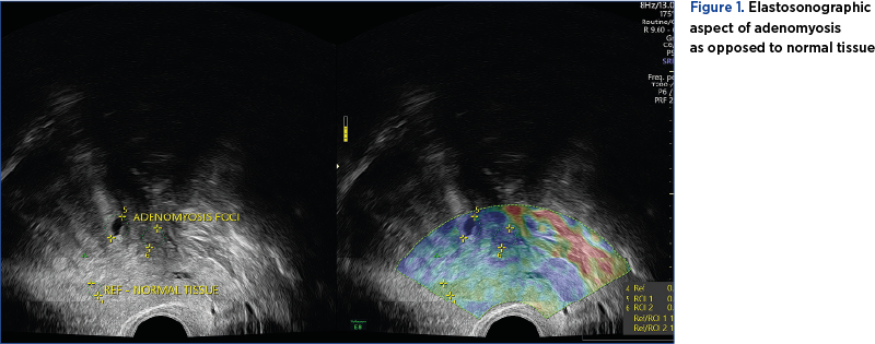
Zhang et al. conducted a prospective study on 34 premenopausal patients, with uterine benign pathology and presenting with pelviabdominal pain and/or metrorrhagia. The quantitative elastography proved to be efficient in detecting adenomyosis foci, by detecting an increase in stiffness tissue as opposed to the healthy surrounding myometrial tissue. However, it failed to differentiate atypical uterine leiomyoma from adenomyosis if the two entities coexisted(19).
In contrast with the aforementioned study, Liu et al. support the use of qualitative elastography in diagnosing adenomyosis, claiming that its accuracy is similar to the one of MRI(20). Furthermore, the study proves that TVUS combined with elastography can differentiate uterine leiomyoma from adenomyosis with an accuracy similar to the one of MRI based on the numeric value of strain ratio (Figure 2)(20).
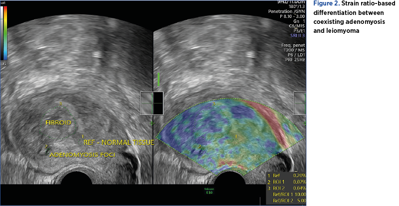
Both techniques require validation in future studies, with strain elastography showing more promising results. The experience of the examiner and the technical equipment also play an important role, both for TVUS and elastography(16).
Biomarkers
The physiopathology and etiology of endometriosis are not yet fully understood. Therefore, various recent studies have tried to elucidate the involvement of various biomarkers in the pathogenesis of this condition. Cytokines, markers of apoptosis, chemotactic molecules, as well as several genes have all proven to play essential roles in the pathways of abnormal implantation of endometrial tissue(21). Among these, interleukin-6 (IL-6), cancer antigen 125 (CA-125) and cancer antigen 199 (CA-199) have been intensly studied as potential biomarkers of endometriosis(22). IL-6, in spite of its involvement in the implantation of endometrial foci, does not increase significantly in patients with endometriosis as opposed to healthy counterparts(23,24).
A meta-analysis performed on small scale studies suggested that serum CA-125 can be used as a reliable, single diagnostic marker in women presenting with symptoms of endometriosis(25). However, other studies underlined its importance only in patients with advanced types of endometriosis(26), and the fluctuation of its levels in relation to phases of the menstrual cycle(27). Similar findings were reported in the case of CA-199, which is also influenced by hormonal secretion and failed to prove itself as a meaningful diagnostic test, when used alone(28).
Circulating endometrium cells (CECs) have benefited from a lot of attention recently, a Chinese study proving that these are detectable in the blood of patients with endometriosis. Their presence highlights the capacity of endometrial tissue to spread through peripheral blood, especially in the case of intrapelvic endometriosis. However, as this study involved a small number of subjects and an even smaller number of patients diagnosed with adenomyosis, further studies on larger populations are necessary for validation(29).
Therefore, as studies of currently trending noninvasive biomarkers have failed to obtain reliable results, the possibility of using these molecules as diagnostic tools for endometriosis is still questionable(22).
MicroRNAs
Ectopic endometrium tissue leads to a chronic inflammatory status, which can be responsible for genic transcription abnormalities and dysregulation of post-translational mechanisms. MicroRNAs, small fragments of non-coding DNA, control the genetic expression through translation inhibition or messenger RNA degradation(30). Therefore, they play an important role in the epigenetic mechanisms responsible for the progesterone-resistant and estrogen-susceptible eutopic endometrial tissue in patients with endometriosis(30). A Chinese study performed on a small-sample size population demonstrated that plasma miR-17-5p, miR-20a and miR-22 are downregulated in patients with stage III and IV endometriosis(31). Another study found an important expression dysregulation of miR-9 and miR-34 miRNA in the eutopic secretory endometrium of women with endometriosis(32). Downregulation of let-7b and miR-135a has also been reported in conjunction with endometriosis, Cho et al. suggesting that these microRNAs have the potential of becoming future noninvasive diagnostic biomarkers. However, the same study recommends the simultaneous use of multiple microRNAs for diagnostic purposes, as well as the necessity of comparing seric and tissular markers of the same type in further studies(33). A Cochrane systematic review recommends the need for further validation of role of microRNAs in further studies as well, highlighting the lack of standardized methods for the detection of various types of these non-coding DNAs, which impacts research reproducibility(28).
Conclusions
The diagnosis of adenomyosis is still challenging, with novel imaging and serological methods emerging as future potential noninvasive assets. The addition of strain elastography seemed to increase the diagnostic accuracy of TVUS, the combined method also being able to distinguish adenomyosis from uterine leiomyoma. Noninvasive biomarkers and microRNAs have showed several intriguing results, but the available data are only based upon small scale population studies. Therefore, further, wider-based population research is necessary to warrant the diagnostic utility of currently studied noninvasive imaging and serological techniques.
elastosonografieadenomiozăbiomarkeri
Marius Geantă: „Solicităm testarea obligatorie a celor 4-5 biomarkeri pentru diagnosticul de precizie în cancerul pulmonar”
Preşedintele Centrului pentru Inovaţie în Medicină, dr. Marius Geantă, a declarat că introducerea testării obligatorie a biomarkerilor pentru diagnosticul de precizie în cancerul pulmonar ar duce la un tratament mult mai ţintit şi ar crea e...