Prezentare de caz: sindrom VACTERL (malformaţii anorectale, cardiace şi renale)
A case report of VACTERL syndrome (anorectal, cardiac and renal malformations)
Abstract
Introduction. VACTERL is a rare condition, named after the spectrum of malformations characterizing it: vertebral/vascular anomalies, anal atresia, cardiac malformations, tracheo-esophageal fistula, renal anomalies, limb anomalies. Case presentation. We report the case of a male infant who presented in our clinic with the suspicion of inborn error of metabolism due to the severe metabolic acidosis and hypoglycemia. He was operated in the second day of life for imperforate anus and rectoperineal fistula. The radiological examination revealed severe bronchopneumonia for which he required antibiotic treatment and electrolyte infusions for the correction of the acidosis. Starting from the anorectal anomalies, we performed imagistic investigations that detected atrial septal defect and unilateral kidney agenesis. Based on the presence of three specific anomalies, we could establish the diagnosis of VACTERL association and we started the supportive treatment. Conclusions. VACTERL association is a complex spectrum of malformations, some of them with high mortality if they are not corrected on the right moment.Keywords
VACTERLimperforate anusatrial septal defectunilateral renal agenesistracheomalaciaRezumat
Introducere. VACTERL este o patologie rară, a cărei denumire este compusă din acronimul malformaţiilor componente: anomalii vertebrale/vasculare, atrezie anală, malformaţii cardiace, fistulă esotraheală, anomalii renale, anomalii ale membrelor. Prezentare de caz. Prezentăm cazul unui sugar în vârstă de 1 lună, care s-a prezentat în serviciul nostru pentru acidoză metabolică severă şi hipoglicemie, ridicând suspiciunea unei boli de metabolism. Pacientul a fost operat în ziua a doua de viaţă pentru imperforaţie anală şi fistulă rectoperineală. Radiografia toracică a decelat bronhopneumonie, pentru care s-a administrat tratament antibiotic, iar pentru corecţia acidozei a necesitat perfuzie cu glucoză şi electroliţi. Având în vedere prezenţa malformaţiei anorectale, am efectuat investigaţii imagistice suplimentare care au decelat un defect septal atrial şi agenezie renală unilaterală. Deoarece pacientul prezenta trei dintre malformaţiile specifice, s-a stabilit diagnosticul de asociere VACTERL. Concluzii. VACTERL este un spectru de malformaţii complexe, care, necorectate în timp util, cresc semnificativ rata de mortalitate a pacienţilor afectaţi.Cuvinte Cheie
VACTERLimperforaţie analădefect septal atrialagenezie renală unilateralătraheomalacieIntroduction
VACTERL association is a rare condition characterized by vertebral/vascular anomalies, anal atresia, cardiac malformations, tracheoesophageal fistula, renal anomalies and limb abnormalities(1,2). It was first described in 1973, with the name VATER, and included vertebral anomalies, anorectal malformations, esophageal atresia, radial anomalies and renal dysplasia(3). After the initial description, other elements were included in this clinical entity, such as vascular anomalies (single umbilical artery) and other limb anomalies (besides radial malformations), and the name changed to VACTERL(1,2). According to EUROCAT (European Surveillance of Congenital Anomalies), the prevalence of VACTERL association was 1 in 20,000 live births in 2012-2016(1).
Recognizing VACTERL cases is essential due to the complexity of component malformations, many of them significantly influencing the long-term prognostic.
Case presentation
We present the case of a 1-month-old male infant who was admitted to our hospital for severe metabolic acidosis (pH 7.21, HCO3 15 mEq/L) and hypoglycemia (40 mg/dL). The patient was born at 39 weeks of gestation by caesarean section. The mother had two previous healthy children, and the pregnancy was not monitored, therefore we do not have any information on the intrauterine evolution of the fetus. His birth weight was 3,300 g, the length was 52 cm, and his Apgar score was 7 due to meconium aspiration and asphyxia. He was operated on the second day of life for anal atresia and rectoperineal fistula. In the postoperative period, he presented repeated episodes of severe metabolic acidosis and hypoglycemia with seizures, receiving treatment with sodium bicarbonate and continuous glucose infusions.
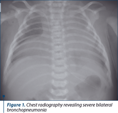
He was admitted to our clinic with the suspicion of an inborn error of metabolism due to the acid-base disorder and electrolyte imbalances. On admission, he weighed 3270 grams, he was lethargic with axial hypotonia and pale skin. The respiratory rate was 60 breaths/minute with subcostal retractions, and the oxygen saturation was 96%. The cardiac frequency was 170 beats/minute, and he presented a second-grade systolic murmur on the entire cardiac area. The abdomen was elastic without hepatosplenomegaly. The anterior fontanelle was normotensive, and he had no other evident anomalies. The laboratory tests revealed metabolic acidosis (pH 7.25, HCO3 16 mEq/L), severe anemia (hemoglobin 7 g/dL) and altered kidney function (creatinine 0.53 mg/dL, urea 60 mg/dL). The inflammatory markers were elevated (CRP 20 mg/dL and procalcitonin 10 ng/mL). The radiological examination revealed bronchopneumonia (Figure 1). We established the diagnosis of respiratory sepsis, and we initiated the intravenous rehydration therapy with sodium bicarbonate supplementation. We started meropenem (60 mg/kg/day) and erythrocyte mass transfusion for the anemia. The evolution was good regarding the acid-base and electrolyte equilibrium, with the normalization of the inflammatory markers. The serum and urinary amino acids and the karyotype were within normal limits.
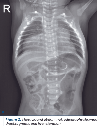
Starting from the anorectal malformation, we investigated for VACTERL association. The cardiology examination with echocardiography revealed an atrial septal defect with nonrestrictive shunt and moderate pulmonary hypertension. The abdominal ultrasound revealed a unique kidney with an abnormal structure. The chest radiography revealed no vertebral anomalies. The abdominal and thoracic computed tomography described diaphragmatic and hepatic elevation, with left kidney agenesis and right ectopic kidney laying on the spine (Figure 2). The bronchoscopy revealed tracheomalacia without tracheoesophageal fistula. The neurological examination detected only axial hypotonia, without other abnormalities.
Based on the presence of three characteristic features (anorectal, cardiac and renal malformations), we could establish the diagnosis of VACTERL association.
Discussion
The purpose of this presentation is to underline the importance of complete head-to-toe investigations in patients with any malformation due to the possibility of complex abnormalities, even though they are not clinically evident.
The diagnosis of VACTERL is mainly made on clinical grounds by the simultaneous presence of the characteristic anomalies after the exclusion of other causes. Any combination is possible, but some authors recognize certain main features, such as tracheoesophageal fistula and anorectal malformations(2). Our patient had anal atresia, atrial septal defect and unilateral renal agenesis.
Due to the imperforate anus and rectoperineal fistula, the meconium could be exteriorized, and he received reconstructive surgery in the first week of life. Anorectal malformations, like imperforate anus/anal atresia, occur in 55-90% of cases. The imperforate anus is usually discovered on the first clinical examination by inspection or lack of meconium elimination(2). The postoperative evolution concerning the reestablishment of the intestinal transit is often more complicated in patients with VACTERL than in those with isolated anorectal malformation(4). The enteral feeding was started early through a nasogastric tube, with good tolerance and the complete reestablishment of the normal intestinal transit in our patient.
The patient had an atrial septal defect with a nonrestrictive shunt and moderate pulmonary hypertension. The cardiac malformations are found in approximately 40-80% of patients and range from severe structural anomalies to mild anatomical defects discovered on routine examinations(2).
An atrial septal defect is one of the most common anomalies, besides ventricular septal defect and the tetralogy of Fallot. The long-term prognosis varies according to the severity of the anomaly, some patients needing only routine clinical follow-up, but others require surgical treatment(5).
Special care is needed for the patients with renal urinary tract involvement, as they are prone to genitourinary infections(2). Our patient had left kidney agenesis, as described on the computed tomography. Genitourinary and renal malformations are found in 50-80% of cases, including unilateral or bilateral renal agenesis (less often), horseshoe kidney and dysplastic/cystic kidneys(2,6). They can be less apparent on the first evaluation, but in some cases, like in our patient, the kidney function is altered, and the imaging studies reveal on first sight the defect.
The bronchoscopy detected tracheomalacia without esophageal atresia or tracheoesophageal fistula. Tracheoesophageal fistula with or without esophageal atresia occurs in 50-80% of cases and is a very severe feature of VACTERL, which requires surgery in the first days of life(2). Some later complications can occur, such as fistula recurrence and gastroesophageal reflux(7).
Although vertebral anomalies are present in approximately 60-80% of VACTERL children and may be associated with rib anomalies, in our patient they were missing. Vertebral malformations include defects like hemivertebrae, dysplastic vertebrae (“butterfly vertebrae”, “wedge vertebrae”), vertebral fusions, and other forms of vertebral dysplasia(2). We will perform periodic follow-up evaluations because some patients present later in life with scoliosis as the first sign if they remain undiagnosed early in life(7).
Other malformations described in VACTERL are the limb malformations, reported in approximately 40-50% of patients, and include thumb hypoplasia/aplasia, polydactyly and lower limb anomalies, with varying range of severity. Facial dysmorphism and neurological impairment are not specific features of VACTERL, and if they are present, other etiologies should be excluded(2).
The role of genetic factors in the etiology of VACTERL is controversial. The cases where gene abnormalities are the cause of the condition were reported in a small number of patients. The most common causes described are mitochondrial dysfunction, pathogenic copy number variations, heterozygous mutations in HOXD13, and heterozygous/hemizygous mutations in ZIC3(2).
Some conditions share multiple elements with VACTERL association (Table 1).

The long-term prognosis is influenced by the severity of the malformations and the success of the surgery treatment. These patients need careful follow-up examinations for the detection of any sequelae that could impact the morbidity. They do not typically present with cognitive impairment.
For the correct management of the patients, it is essential to recognize the life-threatening conditions that must be resolved surgically in the first days of life, such as severe cardiac anomalies, tracheoesophageal fistula and imperforate anus. After managing these conditions, special attention must be paid to the more subtle changes that can affect the later evolution, like vertebral anomalies or renal malformations(2).
Conclusions
VACTERL association is a complex diagnosis and should be looked for if any specific malformations are present in a newborn. The other features may be only subtle manifested, but with severe consequences. Any combination of the malformation spectrum is possible, therefore the cases are very heterogeneous and challenging for any clinician.
Bibliografie
-
Van de Putte R, Van Rooji I, Marcelis C, et al. Spectrum of congenital anomalies among VACTERL cases: a EUROCAT population-based study. Pediatric Research. 2020;87:541-549.
-
Solomon BD. VACTERL/VATER Association. Orphanet Journal of Rare Diseases. 2011;6:56.
-
Quan L, Smith DW. The VATER association. Vertebral defects, anal atresia, T-E fistula with esophageal atresia, radial and renal dysplasia: a spectrum of associated defects. J Pediatr. 1973;82:104-107.
-
Raam MS, Pineda-Alvarez DE, Hadley DW, et al. Long-term outcomes of adults with features of VACTERL association. Eur J Med Genet. 2011;54:34-41.
-
Totonelli G, Catania VD, Morini F, et al. VACTERL association in anorectal malformations: effect on the outcome. Pediatric Surgery International. 2015;31:805-808.
-
Solomon BD, Pineda-Alvarez DE, Raam MS, et al. Analysis of component findings in 79 patients diagnosed with VACTERL association. Am J Med Genet A. 2010;152A:2236-2244.
-
Oral A, Caner I, Yigiter M, et al. Clinical characteristics of neonates with VACTERL association. Pediatrics International. 2012;54:361-364.

Hepatită autoimună de tip 1 asimptomatică cu nivel normal al IgG şi deficit de IgA – prezentare de caz
Alexandra Mititelu, Alina Grama, Mihaela Spîrchez, Claudia Simu, Emilia Pop, Patricia Lorinţiu, Tudor Lucian Pop
Introducere. Hepatita autoimună (HAI) la copii este o boală hepatică cronică inflamatorie, cu un spectru clinic larg, de la o cre...
Spirometric assessment in asthma in children
Bogdan A. Stana, Awwab Shahid
Astmul este o boală importantă, atât la adulţi, cât şi la copii, care conduce frecvent la implicaţii financiare şi de sănătate semnificative la nivel global, afectând peste 300 de milioane de oameni din întreaga lume. Astmul este observat iniţial în copilărie şi se manifestă împreună cu afecţiuni precum f...
Tusea cronică la copil – provocări
Mirela Pavelescu, Irina Dijmărescu, Daniela Păcurar
Introducere. Tusea reprezintă simptomul cel mai frecvent pentru consultaţia la medicul de familie în multe ţări din Europa. Persistenţa ei o perioadă îndelungată afectează calitatea vieţii pacientului. Prezentare de caz. Un adolescent de 13 ani este internat pentru tuse spastică frecventă, emetizantă, diur...
Hepatită autoimună de tip 1 asimptomatică cu nivel normal al IgG şi deficit de IgA – prezentare de caz
Alexandra Mititelu, Alina Grama, Mihaela Spîrchez, Claudia Simu, Emilia Pop, Patricia Lorinţiu, Tudor Lucian Pop
Introducere. Hepatita autoimună (HAI) la copii este o boală hepatică cronică inflamatorie, cu un spectru clinic larg, de la o cre...
Importanţa abordării multidisciplinare a edentaţiei bimaxilare subtotale
Silvia-Izabella Pop, Radu Pop, Andrei Petruţ, Emilia Pop
Reabilitările complexe ale edentaţiilor bimaxilare necesită o planificare atentă şi minuţioasă a detaliilor estetice şi funcţionale....