Technological aspects in the reconstruction of the dental abutment with post and core restoration using the indirect method: modern versus digital
Aspecte tehnologice în refacerea bontului coronar cu ajutorul dispozitivelor coronoradiculare prin metoda indirectă: modern versus digital
Abstract
The loss of a significant amount of dental hard tissues at the coronal level requires the use of a post and core restoration to reconstruct the dental abutment. The role of post and core is to retain the coronal portion of the restoration from the root portion of the tooth and to protect the remaining coronal structures. The post and core can be made classically by the melting-casting process (lost-wax technique) or by the modern version offered by CAD/CAM technology. The latter method, even though it is more expensive, offers multiple advantages, and it is expected to be used more frequently in the future.Keywords
post and corelost wax techniqueCAD/CAMRezumat
Pierderea unei cantităţi semnificative de ţesuturi dure dentare de la nivel coronar impune utilizarea unui dispozitiv coronoradicular pentru reconstruirea bontului dentar coronar. Rolul dispozitivelor coronoradiculare este acela de a retenţiona porţiunea coronară a restaurării de porţiunea radiculară a dintelui şi de a proteja structurile coronare restante. Dispozitivul coronoradicular poate fi realizat în mod clasic prin procedeul de topire-turnare sau prin varianta modernă oferită de tehnologia CAD/CAM. Cea din urmă metodă, chiar dacă este mai costisitoare, oferă multiple avantaje şi este de aşteptat ca în viitor să fie utilizată din ce în ce mai mult.Cuvinte Cheie
dispozitiv coronoradiculartopire-turnareCAD/CAMIntroduction
Restoring the integrity of the teeth represented a human necessity, both for the functionality of the dento-maxillary system and for its esthetic harmony. The loss of a significant amount of dental hard tissues at the coronal level requires the reconstruction of the coronal abutment with the help of post and core. The prosthetic preparation of the tooth is carried out after performing a correct endodontic treatment at the level of the tooth that needs to be prosthetically restored. Thus, post and core are elements contribute to the complete restoration of the coronary abutment, over which the final prosthetic restoration will be made(1-3).
General data
The role of post and core is to retain the coronal portion of the restoration from the root portion of the tooth and to protect the remaining coronal structures. The retention function is dictated by the major indication of the method itself, massive coronary destructions that do not provide conditions for the coronary aggregation of the restoration. The protective function is achieved by transmitting occlusal forces along the axis of the root, in the apical direction, these being distributed in the root and in the supporting marginal periodontium. Specifically, by providing rigidity to the crown device, deformation of the crown edges and cement degradation are prevented(1-3).
Post and core devices extend the longevity of the tooth at the level of the dental arch, giving it a satisfactory prognosis. The objective of prosthetic restoration on endodontically treated teeth should take into account the preservation of the most possible amount of healthy dental substance, this being vital for the prosthetic restoration, considering the existence of a large loss of dental structure(1-3).
The post and core restoration is made so that the healthy tissues at the level of the dental crown are preserved. This restoration is distinct from the final restoration represented by any type of dental crown, which is fabricated, fitted and cemented to the artificial abutment, just like on a natural tooth. The fitting of the dental crown is not conditioned by the fitting of the post and core(1-3).
This paper presents several particular technological aspects in the workflow of some post and core devices, as well as ways to restore the coronal abutments – the classical methods versus digital methods.
Case presentations
The indirect method of obtaining post and core (in the dental laboratory) was chosen, for two distinct technological situations, as follows:
-
The classic method, using lost wax technique, is the method used mostly at the moment in dental practice in Romania, due in particular to the low manufacturing costs.
-
The digital method of indirect fabrication from zirconia through the CAD-CAM technique.
Case 1. Metallic post and core obtained through lost-wax technique (classic method)
A 53-year-old male patient presented in the dental office for a complete restoration of the teeth. This involves, beside prosthetic restoration of the lateral area of the arches, the restoration of the frontal area affected by some major destruction of hard tissues. Clinical and radiological examination revealed that the upper lateral incisor from the first quadrant (1.2) requires a post and core restoration, this being the only solution to keep the tooth on the dental arch. The patient was very satisfied with the fact that the tooth can be saved and maintained at the level of the arch.
Considering the massive coronary destruction, it was concluded that a metallic post and core device, made from Ni-Cr-based alloy, should be used in collaboration with the dental technician, using an indirect method.
It started by performing the endodontic treatment and sealing the root canal properly. After carrying out these mandatory steps, it moved on to the preparation of the canal and the minimum of crown substrate that could be saved.
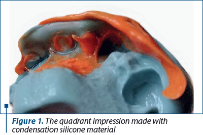
Subsequently, the quadrant impression was made, using two-step impression technique. A condensation silicone material in double consistency was used, the putty as a conformer and the light body at the level of the preparation for a superior fidelity of its details. The impression was then sent to the dental laboratory (Figure 1).
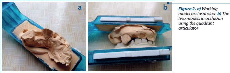
The mandibular and maxillary models were cast using type 4 die stone in a quadrant articulator, for a correct spatial positioning (Figure 2 a and b).
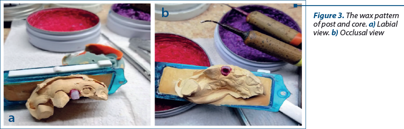
Once the models were obtained, the wax pattern of post and core was created in accordance with the neighboring teeth, the antagonizing teeth, but also with the type of prosthetic restoration that will be made over it (Figure 3 a and b).
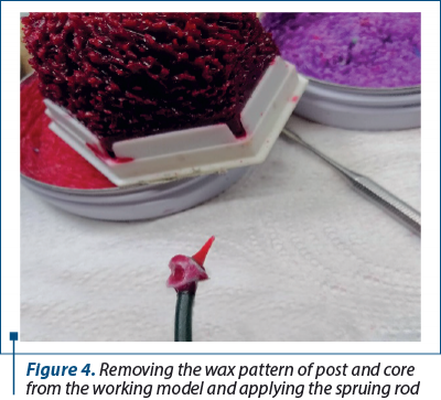
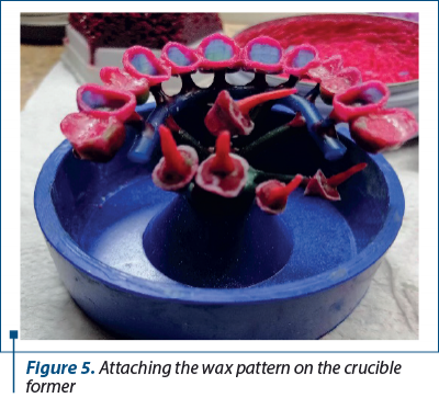
When the wax pattern is ready, a spruing rod is attached, thus preparing the pattern for the investing stage in order to transform it into a metal part. The pattern thus prepared will be applied on the crucible former, the free portion of the casting rod model, and is fixed with wax on the top of the crucible former (Figures 4 and 5).
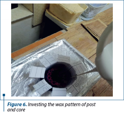
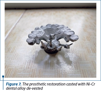
Inside the metallic ring, the investment material was introduced gradually, avoiding the formation of air bubbles that can lead to the appearance of positive defects at the level of the cast parts (Figure 6). After pouring the metal into the mold and cooling the assembly, the metal parts were de-vested and prepared for processing (Figure 7).
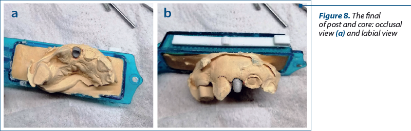
After processing, the post and core device made of Ni-Cr alloy marginally adapted to the level of the preparation on the working model resulted. Afterwards, the post and core device will be sent to the dental office to check the fitting on the tooth and for cementation (Figure 8 a and b).
Case 2. Post and core made of zirconia using CAD/CAM technology (the modern digital method)
A 60-year-old male patient presented to the dental office with a toothache at the level of one of the upper premolars, respectively 2.4. It was found that it had not been properly endodontically treated, so the root canal treatment was redone.
The patient was informed by the dentist about the possibility of making a post and core device in the dental laboratory. The tooth presented a significant destruction, therefore this was the best option for reconstructing the dental abutment with the help of prefabricated post. It was thus concluded that the post and core device made of zirconia was the most suitable for the requirements of the case, as the patient also had the appropriate material possibilities.
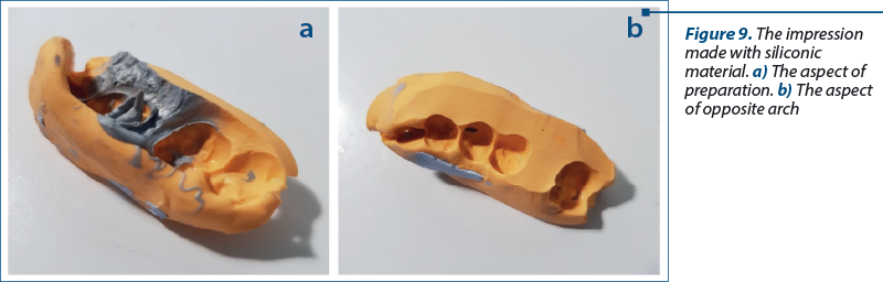
Afterwards, it moved on to the preparation of the root canal and the remaining portion of the crown. A quadrant impression was made using the two step technique with condensation silicone materials. The silicone with putty consistency was used as a conformer in which the light body was added to the level of the preparation for a superior result of the details (Figure 9 a and b).
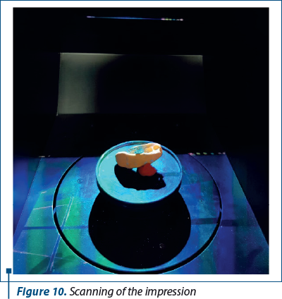
To create the post and core device using CAD/CAM technology, the impression is scanned using a high-precision laboratory scanner. A clear virtual image of the clinical case, including the root preparation, is thus obtained. The scan is performed before casting the two models (maxillary and mandibular) in order to avoid damage of the impression that may cause the appearance of deformations that would lead to modified virtual image compared to reality (Figure 10).
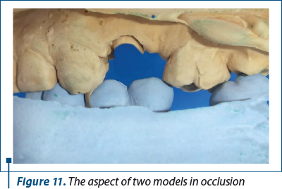
Using the impression of the clinical case, the two models were cast, maxillary with type 4 die stone and mandibular with type 3 die stone, but also their mounting in an occlusal simulator. The physical models were made to check and make the necessary corrections of the post and core device before sending it to the dental office (Figure 11).
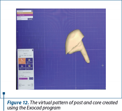
Using the virtual model, the virtual pattern of post and core device was created with the help of the Exocad program. At the root level, a reduction of approximately 2.5% volumetric rate was achieved to create the space needed for the cement used to fix the restoration at the level of the tooth (Figure 12).
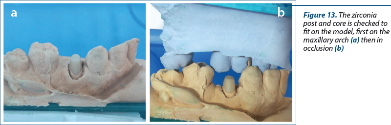
The digital design is then sent to the milling machine to obtain the zirconia post and core device. The milled part is thermally treated in the sintering furnace to stabilize the material. The final shape of the crown-radicular device is checked on the model, aiming at marginal and occlusal adaptation, before sending the restoration to the dental office (Figure 13 a and b).
Discussion
The use of post and core is necessary when the coronal portion of the teeth shows significant destruction and can no longer provide the necessary support for a classic crown restoration(4,5). Classically, the post and core are made of dental alloys, and in the technological process creating the pattern can be done either in the dental office or in the dental laboratory.
Metallic core and post appeared after the development of the melting-casting technology of metal prosthetic parts, in the desire to extend the life of the teeth at the level of the dental arches and thus increase the number of abutment teeth available for creating a fixed prosthetic restoration(6,7). The major disadvantage of these restorations is the unaesthetic appearance they create when used in the frontal area in combination with all-ceramic restorations(8,9). In these situations, even the cements used to retain ceramic restorations cannot favorably mask the gray color given by the dental alloys used(10-12).
The appearance of adhesive cements allowed the use of new categories of materials for the restoration of teeth with significant coronal destruction, namely fiberglass posts. They have a color closer to that of the teeth and allow the use in the frontal areas under ceramic restorations(13,14). The limitations of such materials are related to the destructions that go beyond the tooth cervical line, situations in which the isolation cannot be properly achieved and, implicitly, neither the adhesion of the materials to the dental hard tissues(15,16).
This situation required the development of new materials that allow the production of post and core in the dental laboratory from materials appropriate in color to the dental tissues. Thus, zirconia post and core, which appeared after the development of technologies for obtaining the infrastructure or zirconia crowns, represents a favorable alternative. It solves both the coloristic issue and the one concerning the marginal closure, an issue existing when using fiberglass posts(12,17,18). The working technology does not differ much from that of making zirconia prosthetic restorations, which is why we can expect zirconia post and core to replace classic metallic ones in current practice(4,19-21).
Conclusions
Post and core devices are among the last solutions that can be used to prolong the presence of teeth on the dental arch. There is a very diverse range of post and core devices that adapt to the requirements of each individual case, as well as to the material needs of the patients.
In the last decades, technology has evolved in an accelerated way and, as a result, there are currently very high-performance equipment and technological means. However, we must not forget the early, pioneering periods, in which the technology for performing post and core restorations was based on the lost wax technique. This method represents a safe method, perfected over time and which presents a much lower production cost compared to the new techniques appearing on the market.
Due to these aspects, the classic technology of obtaining post and core by the method of metal casting was practiced and is still practiced in dental laboratories from Romania.
Of course, this technique also has disadvantages, such as the appearance of positive or negative defects on the surface or in the structure of the metal, impurities from the crucible in the mass of the metal, a technique with a high risk of injury, due to the handling of bodies at extremely high temperatures (700-900˚C) etc.
But the technology for creating post and core devices using the CAD-CAM computerized method, although it is much more expensive than the classic technology, represents at the moment a much faster and safer alternative in current dental practice.
Acknowledgement. Viorel Ştefan Perieanu and Manuela Popescu are the corresponding authors and have an equal contribution with the first author.
Conflict of interest: none declared
Financial support: none declared
This work is permanently accessible online free of charge and published under the CC-BY.

Bibliografie
-
Bratu D, Nussbaum R. Bazele clinice şi tehnice ale protezării fixe. Ed. Signata, Timişoara, 2009.
-
Helfer AR, Melnick S, Schilder H. Determination of the moisture content of vital and pulpless teeth. Oral Surg Oral Med Oral Pathol. 1972;34(4):661-670. doi:10.1016/0030-4220(72)90351-9.
-
Oancea L. Consideraţii asupra reabilitării dinţilor devitali cu distrucţii avansate prin reconstituire corono-radiculară. In: Andrei OA, Morar S, Oancea L, Dumitru SG, Actualităţi în asistenţa medical. Vol III. Editura Ars Docendi, Bucureşti, 2013, 217-266.
-
Goracci C, Ferrari M. Current perspectives on post systems: a literature review. Aust Dent J. 2011;56 Suppl 1:77-83. doi:10.1111/j.1834-7819.2010.01298.x.
-
Abou-Rass M. Post and core restoration of endodontically treated teeth. Curr Opin Dent. 1992;2:99-107.
-
Cheung W. A review of the management of endodontically treated teeth. Post, core and the final restoration. J Am Dent Assoc. 2005;136(5):611-619. doi:10.14219/jada.archive.2005.0232.
-
Ricketts DN, Tait CM, Higgins AJ. Post and core systems, refinements to tooth preparation and cementation. Br Dent J. 2005;198(9):533-541. doi:10.1038/sj.bdj.4812300.
-
Lee JD, Khan M, Lee SJ. A Prosthetically Guided Technique for Cast Post-and-Core Fabrication. Compend Contin Educ Dent. 2021;42(9):512-515.
-
Ferrari M, Vichi A, Colt SG, Mason PN. The resin-bonded cast post core: technical preparation and cementation protocols. Pract Periodontics Aesthet Dent. 1997;9(2):233-243.
-
Prakash J, Golgeri MS, Haleem S, et al. A Comparative Study of Success Rates of Post and Core Treated Anterior and Posterior Teeth Using Cast Metal Posts. Cureus. 2022;14(10):e30735. doi:10.7759/cureus.30735.
-
Ge J, Wang XZ, Feng HL. Influence of different post core materials on the color of Empress 2 full ceramic crowns. Chin Med J (Engl). 2006;119(20):1715-1720.
-
Nakamura T, Saito O, Fuyikawa J, Ishigaki S. Influence of abutment substrate and ceramic thickness on the colour of heat-pressed ceramic crowns. J Oral Rehabil. 2002;29(9):805-809. doi:10.1046/j.1365-2842.2002.00919.x.
-
Lamichhane A, Xu C, Zhang FQ. Dental fiber-post resin base material: a review. J Adv Prosthodont. 2014;6(1):60-65. doi:10.4047/jap.2014.6.1.60.
-
Al-Qahtani AS, AlZain SA, AlHamdan EM, et al. A comparative evaluation of the effect of phototherapy of fiber post on its bond strength to dental composite. Photodiagnosis Photodyn Ther. 2018;24:228-231. doi:10.1016/j.pdpdt.2018.08.016.
-
Bitter K, Kielbassa AM. Post-endodontic restorations with adhesively luted fiber-reinforced composite post systems: a review. Am J Dent. 2007;20(6):353-360.
-
Imai D, Mine A, Ezaki R, et al. Does the bonding effectiveness of a fiber post/resin composite benefit from mechanical or chemical treatment? Seven methods for saliva-contaminated surfaces. J Prosthodont Res. 2022;66(2):288-295. doi:10.2186/jpr.JPR_D_21_00015.
-
Suputtamongkol K, Tulapornchai C, Mamani J, Kamchatphai W, Thongpun N. Effect of the shades of background substructures on the overall color of zirconia-based all-ceramic crowns. J Adv Prosthodont. 2013;5(3):319-325. doi:10.4047/jap.2013.5.3.319.
-
Ozkurt Z, Işeri U, Kazazoğlu E. Zirconia ceramic post systems: a literature review and a case report. Dent Mater J. 2010;29(3):233-245.
-
Zou Y, Zhan D, Xiang J, Li L. Clinical research on restorations using CAD/CAM-fabricated monolithic zirconia endocrowns and post and core crowns after up to 5 years. Int J Comput Dent. 2022;25(3):287-294. doi:10.3290/j.ijcd.b2599661.
-
Lee JH. Fabricating a custom zirconia post-and-core without a post-and-core pattern or a scan post. J Prosthet Dent. 2018;120(2):186-189. doi:10.1016/j.prosdent.2017.10.004.
-
Lee JH, Sohn DS, Lee CH. Fabricating a fiber-reinforced post and zirconia core with CAD/CAM technology. J Prosthet Dent. 2014;112(3):683-685. doi:10.1016/j.prosdent.2014.01.015.

Laureaţii primei ediţii a Galei Elitelor Medicale în Otorinolaringologia Românească
Două evenimente de excepţie în domeniul otorinolaringologiei au avut loc, în data de 21 aprilie, la Cluj-Napoca. Este vorba despre Forumul „Progrese şi Excelenţă în Otorinolaringologie” şi Gala Elitelor Medicale în Otorinolaringologia Românească, ambele desfăşurate sub egida Societăţii Române de Foniatrie. ...
Full zirconia single tooth fixed prosthetic restorations obtained through CAD/CAM technology – technological and practical aspects (Part II)
Mihai David, Alexandra Dragomir, Radu Cătălin Costea, Viorel Ştefan Perieanu, Manuela Popescu, Mădălina Violeta Perieanu, Liliana Burlibaşa, Ileana Ionescu, Mihai Burlibaşa
Tehnologia CAD/CAM reprezintă o alternativă inovatoare în protetica dentară, ce asigură o calitate maximă pentru toate tipuri...
Rare clinical features in non-Hodgkin lymphomas of the head and neck
Anca-Ionela Cîrstea, Daniela Vrînceanu, Raluca Grigore, Dumitru Mihai, Maria Dana Gheorghiev, Miruna Bratiloveanu, Nicoleta Măru
Limfomul este cel de-al doilea diagnostic malign, după carcinomul scuamos, cu localizare la nivelul capului şi gâtului. Cea mai frecventă formă de manifestare a limfoamelor din sfera ORL este cea...
Full zirconia single tooth fixed prosthetic restorations obtained through CAD/CAM technology – technological and practical aspects (Part II)
Mihai David, Alexandra Dragomir, Radu Cătălin Costea, Viorel Ştefan Perieanu, Manuela Popescu, Mădălina Violeta Perieanu, Liliana Burlibaşa, Ileana Ionescu, Mihai Burlibaşa
Tehnologia CAD/CAM reprezintă o alternativă inovatoare în protetica dentară, ce asigură o calitate maximă pentru toate tipuri.../38 - Practical aspects regarding manufacturing of complete dentures.webp)
Aspecte practice în confecționarea protezelor totale (Partea I)
Camelia Ionescu, Adelin-Florentin Anca, Viorel Ștefan Perieanu, Florentina Căminișteanu, Mircea Popescu, Ruxandra Stănescu, Cristina Maria Șerbănescu, Mădălina Adriana Malița, Mihai Burlibaşa, Oana Eftene
În ultimii ani, domeniul proteticii dentare a cunoscut o transformare semnificativă, prin integrarea tehnologiilor moderne în pro.../34-Custom impression trays in complete denture technology.webp)